For scientific specimen analysis, you’ll find ImageJ/FIJI, QuPath, and CellProfiler lead the field with robust segmentation algorithms and user-friendly interfaces. Each excels in different applications: ImageJ offers versatility, QuPath specializes in tissue analysis, and CellProfiler handles high-throughput projects. Open-source options provide cost-effective solutions with community support, while commercial alternatives deliver dedicated technical assistance. Your specimen type and research goals will determine which powerful tool will transform your microscopy images into quantifiable insights.
What Digital Image Tools Analyze Scientific Specimens Best?
When selecting digital image analysis tools for scientific specimens, researchers must consider their specific research needs and technical requirements. ImageJ/FIJI stands out as versatile open source software that handles numerous biological image formats while delivering robust quantitative analysis capabilities.
Effective digital image analysis requires tools aligned with your specific research needs and technical demands.
For digital pathology applications, QuPath offers user-friendly tools for tissue segmentation and biomarker assessment without requiring programming expertise.
Cell Profiler’s modular design excels in high-throughput image analysis, processing thousands of images efficiently.
Researchers working with neurons benefit from L-Measure’s specialized neuroanatomical measurements, while collaborative teams should consider Cytomine’s web-based platform for managing large datasets with controlled user access.
The ideal tool ultimately depends on your specimen type, analysis complexity, and whether you need advanced segmentation algorithms or straightforward visualization options.
Key Features of High-Performance Scientific Image Analysis Software
What truly distinguishes high-performance scientific image analysis software from basic image viewers? The answer lies in specialized capabilities designed for biomedical research.
Top-tier tools like ImageJ, CellProfiler, and Fiji offer robust segmentation algorithms that precisely identify cellular and subcellular structures. You’ll find these open source software options provide extensive image processing functionalities for quantification and feature extraction.
Modern solutions integrate deep learning frameworks such as CellPose and U-Net, dramatically improving classification accuracy while reducing manual annotation time. A user-friendly interface remains essential—QuPath exemplifies this by enabling pathologists to analyze whole slide images without extensive programming knowledge.
Collaborative features are increasingly important, with platforms like Cytomine supporting team-based workflows for large-scale studies. These advanced capabilities transform basic visualization into powerful analytical systems that accelerate scientific discovery.
Open Source vs. Commercial Solutions for Microscopy Image Processing
Choosing between open source and commercial solutions for microscopy image processing involves weighing several critical factors beyond mere cost considerations. While ImageJ, Fiji, and QuPath offer flexibility and community-driven improvements without licensing costs, commercial packages provide dedicated support and user-friendly interfaces for less technical users.
| Feature | Open Source | Commercial |
|---|---|---|
| Cost | Free | Subscription/Purchase |
| Support | Community forums | Dedicated technical teams |
| Customization | High flexibility | Limited by licensing |
| Development | Continuous updates | Scheduled release cycles |
You’ll find open source software equally capable for high-throughput image analysis and complex quantitative measurements compared to commercial solutions. The ability to modify code makes open source particularly valuable when you need to tailor analysis workflows to unique experimental designs.
Machine Learning Applications in Advanced Specimen Analysis
Machine learning algorithms like U-Net and CellPose have revolutionized specimen analysis through AI-driven classification methods that efficiently distinguish objects within complex microscopy images.
You’ll find that successful automation in microscopy hinges on robust training datasets, which citizen science initiatives such as ‘Etch a Cell’ help develop for high-throughput analysis applications.
These advanced tools can process large-scale 3D images in real-time, dramatically accelerating your workflow while enhancing image quality through noise reduction and feature enhancement techniques.
AI-Driven Classification Methods
As digital imaging continues to evolve, AI-driven classification methods have revolutionized how scientists analyze biological specimens. You’ll find deep learning algorithms like U-Net and CellPose dramatically improving how microscopy images are segmented and classified.
These AI algorithms learn from large datasets to automatically identify complex biological structures, considerably reducing manual annotation time. You can enhance training data through augmentation techniques like rotations and mirroring, making your classification models more robust across varied imaging conditions.
The real power lies in multi-class classification capabilities, where you can identify multiple specimen types within a single image. By incorporating machine learning into your image analysis workflow, you’ll achieve higher throughput and reproducibility in your research.
This makes AI-driven methods invaluable tools for advancing work in pathology, cell biology, and related scientific fields.
Automation Success Factors
When implementing machine learning for specimen analysis, several key factors determine your success rate. You’ll achieve better results by prioritizing data augmentation techniques like rotations and mirroring, which artificially expand your training datasets and build more robust models for image analysis.
Choose the right tools for your specific tasks—algorithms like U-Net and CellPose excel at segmentation of complex biological structures while efficiently processing large datasets.
Reproducibility improves considerably when you automate analysis workflows, reducing human error and guaranteeing consistent outcomes across multiple specimens.
Don’t overlook accessibility—user-friendly platforms such as ZeroCostDL4Mic provide open-source software options that enable even researchers with minimal coding experience to leverage sophisticated machine learning capabilities.
This democratization of data analysis tools guarantees advanced automation benefits can extend throughout the scientific community.
Multi-Dimensional Data Handling for Complex Biological Structures
Modern multi-dimensional data handling tools now offer you powerful options for 3D cell reconstruction, including ImageJ’s volumetric rendering and VIAS’s confocal stack integration capabilities.
You’ll find multi-scale analysis methods particularly valuable when examining structures that span different organizational levels, with QuPath excelling at tissue-wide quantification while CellProfiler focuses on cellular details.
Time-series visualization tools such as Fiji’s hyperstacks let you track dynamic biological processes across temporal dimensions, revealing patterns that would remain hidden in static images.
3D Cell Reconstruction Options
The analysis of complex biological structures demands sophisticated digital tools capable of handling multi-dimensional data with precision and flexibility. When you’re working with d cell reconstruction tools, your options include specialized software like L-measure, which quantifies morphological features of neurons in both 2D and 3D images.
For more extensive reconstruction needs, consider:
- The Volume Integration and Alignment System (VIAS), which unifies multiple confocal microscopy image stacks into interactive 3D datasets.
- CellProfiler’s custom pipelines that extract quantitative measurements from large datasets efficiently.
- QuPath’s user-friendly digital pathology image analysis tools for tissue segmentation.
Advanced deep learning models like U-Net and CellPose have revolutionized segmentation tasks, delivering enhanced accuracy when analyzing intricate biological specimens in multidimensional datasets.
Multi-Scale Analysis Methods
Multi-scale analysis methods build upon 3D reconstruction approaches by expanding your analytical capabilities to navigate across dimensional boundaries.
When handling complex biological structures, you’ll need tools like QuPath and Cell Profiler that provide frameworks for automated analysis of large image datasets.
For effective multi-scale analysis, implement image segmentation and feature extraction techniques to isolate specific components within larger biological systems. These approaches help you quantify structural features at various levels—from cellular organization to complete tissue architecture.
Deep learning algorithms enhance your analytical power tremendously. Tools like U-Net and StarDist improve segmentation accuracy in high-dimensional imaging data, identifying intricate patterns that traditional methods might miss.
When combined with advanced imaging techniques like intravital microscopy, you’ll gain unprecedented insights into dynamic biological processes in their natural context, revealing how structures function across multiple scales simultaneously.
Time-Series Visualization Tools
Analyzing dynamic cellular behaviors demands specialized time-series visualization tools that transform complex temporal data into interpretable patterns. When you’re tracking morphological changes across multiple time points, these tools integrate seamlessly with platforms like ImageJ and QuPath to handle large datasets efficiently.
- Watch cells migrate and interact in real-time as advanced algorithms capture subtle changes in cellular features and membrane dynamics.
- Manipulate your visualization with interactive features that let you rotate, zoom, and animate complex biological structures to reveal hidden temporal patterns.
- Process multi-dimensional data without bottlenecks by employing sufficient computational resources that manage high-frequency imaging output.
The best time-series tools balance sophisticated analysis capabilities with intuitive interfaces, allowing you to extract meaningful insights from temporal dynamics without getting overwhelmed by technical complexity.
Quantitative Analysis Tools for Cell Morphology and Distribution
While visual assessment provides qualitative insights, modern digital analysis tools offer researchers unprecedented capabilities to extract precise measurements from cellular images.
CellProfiler and QuPath stand out as powerful quantitative analysis tools that enable you to measure cell area, shape, and distribution with remarkable accuracy.
You’ll find Cell Profiler Analyst particularly valuable if you’re new to programming, as it allows efficient analysis of large datasets without coding requirements.
For neuronal studies, L-Measure delivers detailed quantification of dendritic structures and cell morphology.
When working with large-format images, Orbit’s automated capabilities support high-throughput analysis across extensive datasets.
Additionally, ImageJ/FIJI excels at measuring fluorescence intensity and protein co-localization, giving you extensive tools to characterize cell morphology and distribution in your scientific specimens.
Integration Capabilities With Microscopy Hardware Systems
Successful scientific imaging depends on seamless communication between software tools and microscopy hardware systems. When selecting digital image analysis platforms for your biomedical image analysis workflow, consider how they integrate with your existing equipment.
Open source software like Fiji and ImageJ offers extensive compatibility through plugins that support various microscope brands and imaging modalities.
- Micro-Manager enables real-time interaction with microscopy hardware, allowing direct control of image acquisition while simultaneously processing your data.
- Web-based platforms like Cytomine optimize collaboration by connecting multiple users to the same microscopy setup, particularly valuable for analyzing large datasets.
- Software such as QuPath and Orbit support both local and remote image loading, making them ideal for whole slide imaging systems and digital pathology scanners.
Batch Processing Solutions for Large-Scale Research Projects
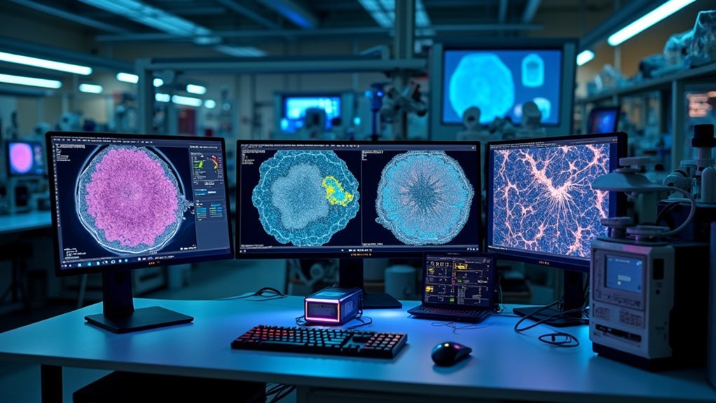
As research projects expand in scope and complexity, handling thousands of scientific images manually becomes virtually impossible. Batch processing solutions like CellProfiler deliver automated analysis capabilities that drastically reduce processing time for cellular and subcellular studies.
You’ll find specialized image analysis software tools such as QuPath and Orbit particularly valuable for pathology research, as they enable pipeline creation for whole-slide image datasets.
ImageJ/Fiji’s batch capabilities guarantee consistency and reliability by applying identical protocols across multiple images. For collaborative projects, Cytomine’s web-based platform facilitates simultaneous processing and annotation.
Beyond efficiency, these automated approaches minimize human error when managing large volumes of data—a critical factor in achieving high-quality results in large-scale research projects where precision can’t be compromised.
Frequently Asked Questions
Which Software Is Used to Analyse an Image?
You can use ImageJ/FIJI, QuPath, CellProfiler, Orbit, or Cytomine to analyze images. Each offers different strengths—ImageJ for versatility, QuPath for pathology, CellProfiler for high-throughput, and Cytomine for collaboration.
What Are the Two Major Types of Image Analysis?
The two major types of image analysis are quantitative and qualitative. You’ll use quantitative analysis for measurable data extraction and statistical evaluation, while qualitative analysis helps you interpret visual characteristics and patterns subjectively.
Which Software Is Used for Digital Image Processing?
For digital image processing, you’ll find ImageJ/FIJI, CellProfiler, and QuPath are widely used. You can also utilize deep learning tools like U-Net and CellPose for advanced segmentation of your scientific images.
What Is the Open Source Software for Digital Pathology Image Analysis?
For digital pathology image analysis, you’ll find several powerful open source options: QuPath for whole slide images, ImageJ/Fiji with its extensive plugins, and CellProfiler for high-throughput cellular analysis. They’re all continuously improved through community contributions.
In Summary
You’ll find no single “best” imaging tool for all scientific specimens. Instead, choose software that matches your specific research needs, budget constraints, and technical expertise. Consider tools with robust segmentation capabilities, machine learning integration, and batch processing for large datasets. Whether you select open-source solutions like ImageJ or commercial packages like Imaris, prioritize tools that efficiently handle your specimen type and research questions.



Leave a Reply