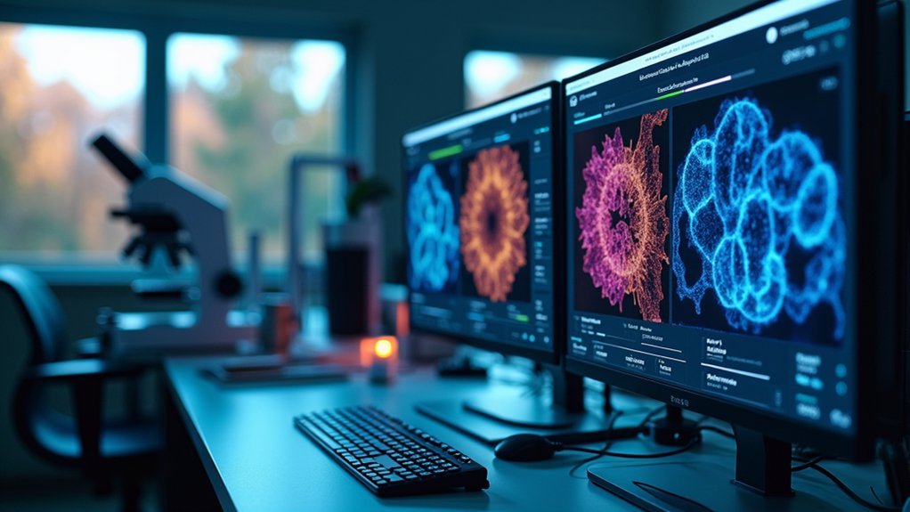
The 7 best image contrast enhancement tools include TopazLabs for AI-driven microscopy enhancement, Adobe’s Scientific Imaging Suite for deconvolution, AVCLabs Photo AI for staining optimization, ImageJ/Fiji for histology samples, batch processing solutions like Photo Enhancer AI, non-destructive editors like Photoshop and Luminar AI, and real-time enhancement software for live microscopy. Each tool offers unique capabilities to improve visibility while preserving scientific integrity in your research images. Discover which solution best fits your specific imaging challenges.
TopazLabs: The Ultimate Contrast Enhancement Solution for Microscopy

When examining microscopic specimens, image clarity can make or break your research findings. TopazLabs offers a powerful solution with its AI-driven image enhancement technology specifically beneficial for microscopy applications.
The software’s Supercontrast tool lets you refine tonal contrasts, bringing out subtle details in your microscopic samples that might otherwise remain hidden.
You’ll appreciate the intuitive interface featuring auto-enhancement options that work effectively regardless of your technical expertise level.
TopazLabs supports essential file formats including TIFF, JPG, and PNG, ensuring seamless integration with standard microscopy imaging systems.
When you’re working with multiple samples, the batch processing capability saves valuable time by enhancing numerous images simultaneously, streamlining your workflow and maintaining consistent quality across your entire dataset.
Adobe’s Scientific Imaging Tools for Microscope Photography
Adobe’s scientific imaging tools offer powerful deconvolution features that you’ll find invaluable for separating overlapping structures in microscope images.
You can utilize their extensive Scientific Image Analysis Suite to measure, quantify, and extract meaningful data from your microscopy specimens.
These specialized tools integrate seamlessly with your existing workflow, allowing for swift analysis while maintaining scientific accuracy and reproducibility.
Advanced Deconvolution Features
Although not widely known outside scientific circles, the advanced deconvolution capabilities in Adobe’s scientific imaging toolkit represent a quantum leap for microscope photography.
These powerful algorithms effectively reverse optical aberrations, dramatically enhancing image quality by reducing blur from lens imperfections or sample movement.
You’ll appreciate the flexibility to select between Wiener or Richardson-Lucy deconvolution methods, optimizing results for your specific imaging conditions.
The software supports high-resolution image formats commonly used in research, ensuring seamless compatibility with various microscopy techniques.
What truly sets these tools apart is their user-friendly accessibility.
You don’t need extensive technical knowledge to perform complex image analyses – the intuitive interface lets you visualize fine microscopic details with remarkable clarity, elevating the quality of your scientific publications.
Scientific Image Analysis Suite
Researchers worldwide count on Adobe’s Scientific Image Analysis Suite as their go-to solution for microscope photography enhancement. The suite delivers advanced photo editing tools specifically designed for scientific imaging that help you enhance images while maintaining data integrity through non-destructive editing techniques.
- Precise histogram adjustments let you fine-tune contrast enhancement for ideal image quality and accurate interpretation.
- Advanced filters provide sophisticated manipulation options tailored to microscopy visualization needs.
- Batch processing capabilities enable you to efficiently process multiple images simultaneously for large research projects.
- Extensive file format support guarantees seamless integration with your existing laboratory workflows.
You’ll appreciate how these tools balance powerful contrast enhancement capabilities with scientific accuracy, making Adobe’s suite an essential component of modern research imaging technology.
AVCLabs Photo AI: Deep Learning for Specimen Staining Optimization

When examining microscopic specimens, image quality can make or break your analysis. AVCLabs Photo Enhancer leverages deep learning algorithms to optimize specimen staining in your images, revealing details that might otherwise remain hidden.
Image clarity reveals microscopic truths that conventional analysis might miss, making AI enhancement essential for modern specimen examination.
You’ll appreciate the automatic quality improvement features that highlight vital elements while preserving the natural-looking enhancements of your samples. The software minimizes artifacts and maintains image integrity, essential for accurate scientific interpretation.
If you’re processing multiple slides, the batch processing capability lets you enhance numerous images simultaneously, streamlining your research workflow.
Compatible with TIFF, JPG, and PNG formats, you’ll find it adaptable to various imaging protocols. The skin retouching and enhancement tools further help emphasize important features in your stained specimens for more effective analysis.
Specialized Contrast Enhancement Techniques for Histology Samples
You’ll need specialized tools like ImageJ or Fiji when working with histological samples to truly visualize subtle variations in tissue staining.
These platforms offer advanced staining visualization capabilities through histogram equalization and adaptive contrast techniques that highlight cellular structures while preserving diagnostic integrity.
For complex specimens requiring multi-channel adjustment, AI-powered solutions like Topaz Labs can simultaneously enhance different staining channels while reducing noise through intelligent deconvolution algorithms.
Advanced Staining Visualization Tools
Although general-purpose image editors can handle basic contrast adjustments, histology samples often require specialized solutions that understand the unique challenges of biological imaging.
Advanced staining visualization tools like MorpholibJ and CLIJ are designed to enhance contrast in histology images using algorithms specifically tailored for biological specimens.
- Deconvolution techniques separate overlapping stained tissues, clarifying boundaries and improving overall image quality
- AI-powered plugins automatically optimize contrast while preserving the quantitative accuracy of your pixel values
- Gaussian blurring and median filters remove random noise without sacrificing important structural details
- Tools streamline documentation of preprocessing steps, ensuring reproducibility in your research
When working with delicate histological samples, these specialized tools provide precision that general image editors simply can’t match.
Multi-Channel Adjustment Solutions
Histology samples often reveal their secrets only when multiple imaging channels work in harmony to highlight different cellular components. Multi-channel adjustment solutions employ specialized algorithms that analyze multiple data channels simultaneously, greatly enhancing contrast in complex tissue samples.
When you’re working with overlapping fluorescence signals, spectral unmixing techniques can differentiate these signals with precision. Advanced software tools applying histogram equalization across various channels optimize brightness and contrast, making cellular structures pop with clarity.
You’ll find adaptive contrast enhancement methods particularly valuable as they preserve histological details while amplifying specific features like nuclei or cytoplasm.
For consistency in your research, consider AI-driven tools that automate the enhancement process, reducing manual adjustments and boosting reproducibility in analysis. These technologies transform how you visualize and interpret histological data.
Batch Processing Tools for High-Volume Microscope Image Analysis

Three key challenges face researchers working with microscope images: volume, consistency, and time.
Batch processing tools offer an elegant solution, allowing you to enhance contrast across hundreds of images simultaneously while maintaining quality standards.
Software options like AVCLabs Photo Enhancer AI, TopazLabs, and Fotor leverage advanced AI algorithms to streamline your workflow:
- Process entire experimental datasets overnight while you focus on analysis
- Apply consistent enhancement parameters across all images regardless of file formats
- Preserve vital scientific details while improving visibility of cellular structures
- Handle TIFF, JPG, and other formats without quality degradation
Non-Destructive Contrast Adjustment for Scientific Documentation
When documenting scientific findings, preserving the integrity of your original image data is non-negotiable. Non-destructive contrast adjustment techniques allow you to enhance visual distinction without compromising your raw data’s integrity.
Modern image editing tools like Adobe Photoshop and Luminar AI offer adjustment layers that let you modify and revert changes at any time. When enhancing scientific images, you’ll need to maintain balance to avoid oversaturating and losing critical information.
| Software | Features | Scientific Benefits |
|---|---|---|
| Adobe Photoshop | Highlights, midtones, shadows sliders | Precise control without data loss |
| Luminar AI | Non-destructive adjustment layers | Original pixel preservation |
| ImageJ | Documentation of adjustments | Enhanced reproducibility |
These non-destructive methods support scientific documentation by enabling consistent adjustments across multiple images while maintaining a clear record of modifications for reproducibility in your research.
Real-Time Contrast Enhancement for Live Microscopy Applications

Live microscopy demands instant image optimization to capture fleeting cellular interactions that might otherwise go undetected.
Modern software tools now offer real-time contrast enhancement capabilities that transform your imaging workflow through advanced algorithms and AI-driven denoising techniques.
When selecting live microscopy software, look for these essential features:
- Adjustable contrast and brightness sliders with immediate visual feedback
- High dynamic range (HDR) imaging that reveals details in both light and dark regions simultaneously
- AI-powered denoising that separates signal from noise while preserving structural integrity
- Automated contrast adjustments that adapt to changing sample conditions without manual intervention
These capabilities guarantee superior image quality during experiments, eliminating the need for extensive post-processing and allowing you to focus on scientific discovery rather than technical adjustments.
Frequently Asked Questions
Which Tool Is Used to Increase Contrast of Image?
You can increase image contrast using Adobe Express, TopazLabs’ Supercontrast, Fotor’s one-tap enhancement, Luminar AI’s algorithms, or AVCLabs and FliFlik’s extensive tools. Each offers unique features for enhancing your photos’ tonal range.
What Is the Most Accurate Photo Enhancer?
Topaz Labs stands out as the most accurate photo enhancer. You’ll appreciate its advanced noise reduction and image sharpening capabilities, making it an excellent choice for your professional photography enhancement needs.
What Is the Best Tool to Increase Image Resolution?
For increasing image resolution, Topaz Labs’ Gigapixel AI is your best choice. You’ll get up to 600% upscaling while maintaining impressive sharpness and detail through its advanced AI algorithms.
Can Chatgpt Enhance Images?
No, ChatGPT can’t enhance images as it’s a text-only AI. You’ll need dedicated software like Adobe Express, Topaz Labs, or Luminar AI for contrast and resolution improvements in your photos.
In Summary
You’ve got powerful options for enhancing your microscopy images. Whether you’re using Topaz for ultimate clarity, Adobe’s scientific tools, or AI-driven solutions like AVCLabs, you’ll find the right software for your needs. Don’t forget specialized histology tools, batch processing for high volumes, non-destructive workflows, and real-time solutions. Choose the tool that matches your scientific imaging requirements for consistently professional results.


Leave a Reply