Achieve crystal-clear scientific photos by mastering diffuse lighting techniques that eliminate harsh shadows and minimize glare. Use large light sources positioned at 45-degree angles from your specimen, and opt for dome lights with reflective surfaces or flat diffusers in tight spaces. Adjust color temperature between 5000K-6500K for accurate representation, and control intensity based on specimen opacity. These fundamentals of diffuse illumination will transform your scientific documentation into publication-quality images with every detail precisely captured.
Numeric List of 9 Second-Level Headings
Nine essential topics comprise the framework for mastering diffuse lighting in scientific photography. These second-level headings provide a thorough roadmap for achieving ideal results:
Mastering diffuse lighting requires understanding nine critical elements that form the foundation for exceptional scientific image quality.
- Understanding Diffuse Light Properties
- Selecting Appropriate Light Sources and Sizes
- Positioning Techniques for Even Illumination
- Managing Shadow Elimination Strategies
- Wavelength Selection for Maximum Detail
- Edge-lit vs. Flat Diffuse Systems
- Compatibility Assessment with Existing Equipment
- Environmental Adaptations for Lighting Setups
- Testing and Optimization Protocols
Each heading addresses critical components of diffuse lighting implementation.
You’ll progress from basic principles to advanced applications, ensuring your scientific photos reveal intricate details with exceptional clarity.
Understanding the Science Behind Diffuse Illumination
Diffuse illumination works by scattering light particles across multiple angles, creating an even distribution that reduces harsh shadows on your scientific specimens.
You’ll find that larger light sources relative to your subject produce softer shifts between light and dark areas, effectively eliminating distracting shadows that might obscure important details.
When you position diffusers between your light source and subject, you’re forcing light rays to travel through translucent materials that break up direct beams into countless redirected paths, resulting in the gentle, uniform lighting essential for accurate scientific documentation.
Light Scattering Principles
While photographers often use diffuse lighting intuitively, understanding the physical principles behind light scattering can greatly improve your scientific imaging results.
When light from your sources encounters particles or irregularities in a medium, it deviates from its straight path, creating diffuse illumination. Rayleigh scattering explains why blue light scatters more than red, affecting the color quality of your diffuse lights. Mie scattering, involving larger particles, creates the even illumination essential for detailed scientific imaging.
- Shorter wavelengths scatter more intensely, influencing color accuracy in microscopy
- Uniform scattering reduces contrast and highlights, revealing fine specimen details
- Quantifying light uniformity helps optimize your imaging setup
- Different diffuse light sources create varying scattering patterns
- The angle of scattered light affects the visibility of surface textures and structures
Shadow Elimination Methods
Because shadows can obscure vital details in scientific specimens, mastering effective elimination techniques is essential for accurate documentation and analysis.
To minimize shadows in your images, utilize translucent diffusers or softboxes that scatter light evenly across your subject’s surface. These materials transform harsh direct light into soft, uniform illumination that reveals subtle details.
Remember that larger light sources relative to your subject produce softer shadows. Consider edge-lit flat diffuse lights for high-intensity, shadow-free illumination of complex specimens. They’re particularly effective at dispersing light evenly across the entire inspection area.
For best results, you’ll need to carefully adjust the distance and angle of your light sources. Pay attention to how they interact with your specimen’s surface texture—this relationship is vital for effectively eliminating shadows through diffuse lighting techniques.
Essential Equipment for Optimal Diffuse Lighting

To achieve professional-quality scientific photography, you’ll need specialized lighting equipment that eliminates harsh shadows and reduces unwanted reflections. Diffuse LED illuminators produce a gentle, even light that creates the ideal “cloudy day” effect for your specimens.
By selecting the appropriate intensity of light, you can guarantee proper exposure without introducing heat that might damage delicate samples.
- Axial illuminators provide uniform lighting across your entire setup
- Color options (white, red, blue, green) enhance contrast for specific applications
- High-intensity models excel when inspecting reflective or curved surfaces
- Custom-sized flat diffuse lights work effectively at larger distances
- Edge-lit panels with high-current LEDs eliminate hotspots for superior image quality
Comparing Dome Lights vs. Flat Diffusers for Microscopy
When selecting between dome lights and flat diffusers for your microscopy setup, you’ll notice substantial differences in light distribution patterns. Domes provide non-directional illumination ideal for curved surfaces, while flat diffusers create a more uniform “cloudy-day” effect at greater working distances.
You can virtually eliminate specimen shadows with either option, though dome lights excel with highly reflective samples when positioned at close range and oversized by approximately 50% relative to your specimen.
Your available space will also influence this decision, as flat diffusers offer customizable dimensions and placement flexibility, whereas dome lights maintain fixed geometries that may present installation challenges in confined microscopy workstations.
Light Distribution Patterns
The choice between dome lights and flat diffusers represents a critical decision in microscopy illumination strategy.
When selecting your light source, consider how distribution patterns affect image quality. Dome lights provide soft, non-directional illumination ideal for curved surfaces, while flat diffusers create a “Cloudy Day” effect at larger working distances.
For best results, your light source dimensions should exceed your specimen size by approximately 50%, guaranteeing even coverage without shadowing.
- Dome lights minimize shadows, critical for highly detailed inspections
- Flat Domes achieve uniform light distribution at specific working distances
- Edge-lit flat diffusers deliver up to 7,000 Lux intensity for detailed imaging
- Custom sizing options allow for application-specific tailoring
- The 50% size rule guarantees complete illumination coverage across your specimen
Specimen Shadow Elimination
Shadows lurking across your specimens can obscure critical details and compromise scientific analysis, making shadow elimination a fundamental concern in microscopy imaging. When choosing between dome lights and flat diffusers, consider your specific application needs.
| Feature | Dome Lights | Flat Diffusers |
|---|---|---|
| Working Distance | Excels at close range | Better for larger distances |
| Light Distribution | Non-directional, curved coverage | “Cloudy Day” uniform effect |
| Best Applications | Highly reflective surfaces | Complex geometries, larger specimens |
| Customization | Adjustable size & wavelength | Adjustable size & wavelength |
Dome lights provide superior shadow elimination on curved surfaces by delivering broad, non-directional illumination from multiple angles. For larger specimens, you’ll find flat diffusers more effective, as they distribute light evenly across wider areas. Both options offer high-intensity configurations for enhanced contrast, ensuring you’ll capture the precise details required for accurate scientific documentation.
Space Constraints Analysis
Despite their exceptional illumination qualities, dome lights and flat diffusers present distinct challenges when integrated into space-limited microscopy environments.
When shipping your equipment between labs, you’ll need to evaluate which lighting solution provides the correct balance between illumination quality and spatial practicality.
- Dome lights require 50% more space than your specimen but deliver superior 360-degree shadow elimination
- Flat diffusers offer a lower profile solution, ideal for cramped microscopy setups
- Assess vertical clearance—dome fixtures need height while diffusers need width
- Both options can be customized in size and wavelength to fit your specific constraints
- In extremely tight spaces, a flat diffuser might be your only option despite the dome’s illumination advantages
Your choice ultimately depends on weighing illumination quality against the practical space limitations of your microscopy setup.
Eliminating Glare on Highly Reflective Specimens
When photographing highly reflective specimens, unwanted glare can greatly compromise image quality and obscure essential details. To eliminate glare, position your light source at an angle to the specimen rather than directly above it. This simple adjustment prevents direct reflections from reaching your camera.
Angle your lights strategically to prevent reflections from ruining detail in shiny specimen photography.
Utilize diffuse lighting techniques with machine vision dome lights to provide even illumination across your subject. These specialized lights distribute illumination uniformly, minimizing harsh highlights on reflective surfaces.
For particularly challenging specimens, combine polarized filters with your diffuse lighting setup. This powerful combination further reduces unwanted reflections while maintaining accurate color representation.
Experiment with flat diffuse lights or edge-lit models in different wavelengths to find the best setup for your specific material.
White light works well for many applications, but certain colored lights might better eliminate glare on your particular specimens.
Adjusting Light Intensity for Various Specimen Types
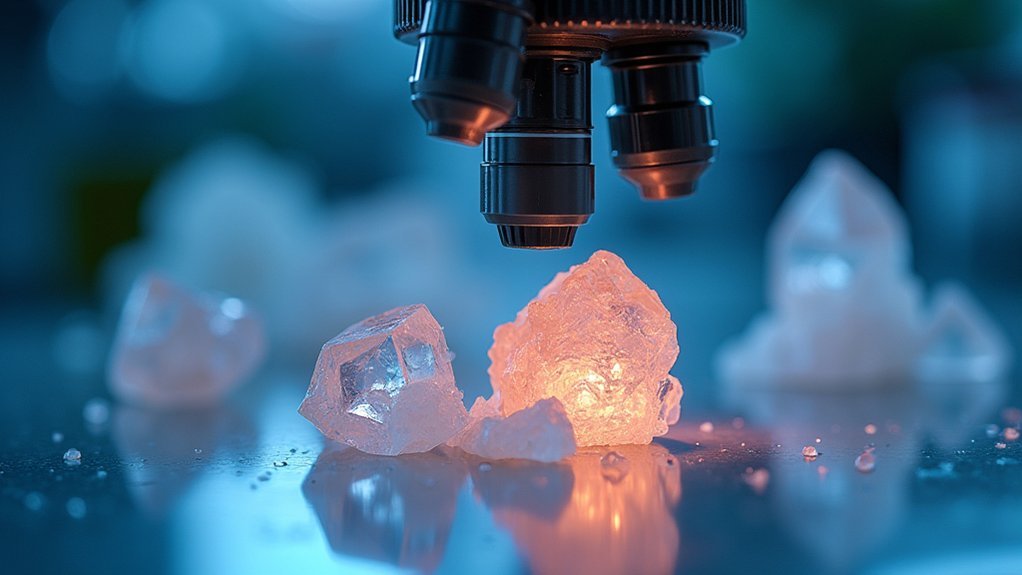
Beyond eliminating glare, mastering light intensity represents the next key challenge in scientific photography. When adjusting light intensity for accurate images, consider the opacity of your specimen. Opaque materials require higher intensity, while translucent or delicate specimens need gentler illumination to prevent damage and capture subtle details.
- Use LED diffuse lights with adjustable settings for precise control
- Decrease intensity for reflective specimens like metals to enhance surface details
- Increase brightness gradually for dense biological or geological samples
- Monitor exposure levels continuously as specimen textures and colors influence lighting needs
- Apply uniform illumination through diffuse lighting to minimize shadows that obscure important features
Remember to make real-time adjustments during your imaging session—lighting that works perfectly for one specimen may completely wash out another.
Color Temperature Considerations for Scientific Accuracy
Accurate color reproduction stands at the core of reliable scientific documentation, with color temperature playing an essential role in this process. When capturing scientific images, aim for lighting between 5000K and 6500K—this daylight-equivalent range guarantees accurate representation of your specimens’ true colors.
Lower temperatures around 3000K produce warm, yellowish tones, while higher settings above 6000K create cooler, bluish casts. Either extreme can distort your sample’s appearance and lead to misinterpretation of results.
For consistent color fidelity, always calibrate your camera’s white balance settings before shooting. Consider using specialized LED lights with adjustable color temperature or appropriate filters to maintain uniform lighting conditions.
Remember that consistency across all your imaging sessions is essential—maintaining the same color temperature standards will greatly enhance the reliability and reproducibility of your scientific observations.
Positioning Techniques for Shadow-Free Documentation

The strategic positioning of diffuse lighting represents one of the most vital factors in capturing shadow-free scientific photographs. When you place your light source at a 45-degree angle to your subject, you’ll maximize soft illumination while minimizing harsh shadows that can obscure important details.
For best results, verify your lighting equipment is approximately 50% larger than your subject. This size difference creates even illumination across curved or reflective surfaces, which is important for accurate documentation.
- Position lights closer to the subject for enhanced diffusion and uniform distribution
- Use large softboxes or reflectors to increase the relative size of your light source
- Experiment with the 45-degree angle for ideal shadow reduction
- Consider multiple diffuse lighting sources to eliminate remaining shadows
- Adjust distance between illumination source and subject for best softness
Troubleshooting Common Diffuse Lighting Challenges
Despite careful setup, you’ll likely encounter several challenges when implementing diffuse lighting for scientific photography. When your images show harsh shadows or uneven illumination, check if your light source is proportionally sized to your subject—larger subjects require larger diffusers for consistent results.
If you’re still seeing unwanted glare on reflective specimens, adjust the angle of your light source or reposition your diffuser. Remember that the diffuser-to-subject distance greatly impacts softness; move it closer for gentler lighting or farther for more definition.
Watch for deteriorating diffusing materials, as worn softboxes or umbrellas can create inconsistent lighting patterns.
Combine natural and artificial diffusers strategically when a single solution isn’t providing adequate diffusion, especially for challenging subjects with both reflective and matte surfaces.
Frequently Asked Questions
What Do Photographers Use to Diffuse Light?
You’ll use natural diffusers like clouds or fog, and artificial tools such as softboxes, umbrellas, and diffuser panels. You can also bounce flash off walls and ceilings for softer, more flattering light.
What Paper Is Best for Diffusing Light?
For diffusing light, you’ll find translucent tracing paper works excellently for cost-effective solutions. If you’re seeking premium results, specialty polycarbonate or acrylic diffusion films provide superior performance with LEDs, eliminating harsh hot spots.
What Is a Soft and Diffused Lighting Technique Used to Create Flattering Portraits?
You’ll achieve flattering portraits with soft, diffused lighting by using large softboxes positioned close to your subject. This technique wraps gentle light around facial features, minimizing harsh shadows and creating a natural appearance.
What Are the Different Types of Diffused Lighting?
You’ll encounter various diffused lighting types including natural diffusers (clouds, fog), artificial diffusers (softboxes, umbrellas), dome lights, flat diffuse lights, and edge-lit flat diffusers—each softening light to reduce harsh shadows and glare.
In Summary
You’ve now mastered diffuse lighting techniques for scientific photography. By understanding light behavior, selecting appropriate equipment, and positioning your setup correctly, you’ll consistently capture crystal-clear images. Remember, proper diffusion eliminates distracting reflections, reveals true specimen details, and guarantees reproducible results. Keep experimenting with these techniques to find the best lighting configuration for your specific specimens and research needs.


Leave a Reply