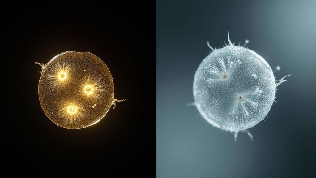
Dark field microscopy creates bright specimens against dark backgrounds by capturing only scattered light, ideal for visualizing external structures of small organisms. Phase contrast converts invisible phase shifts into visible amplitude differences, revealing internal cellular details without staining. You’ll need specialized condensers for dark field, while phase contrast requires precise phase rings in both condenser and objective. Choose dark field for motile microorganisms and phase contrast for cellular morphology. The right technique depends on your specific specimen and research goals.
Optical Principles Behind Dark Field and Phase Contrast
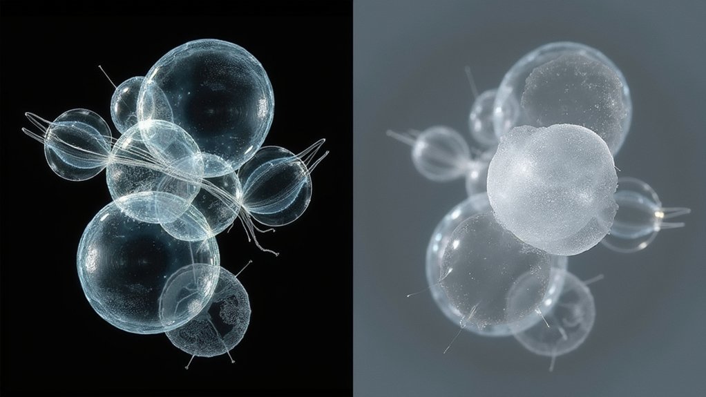
While both techniques revolutionized microscopy for unstained specimens, dark field and phase contrast operate on fundamentally different optical principles.
In dark field microscopy, a specialized condenser blocks direct light rays while allowing scattered light to reach the specimen. This creates a striking contrast image where your biological samples appear bright against a dark background, as light reflects off their surfaces.
You’ll see fine details in small organisms with remarkable clarity.
Phase contrast microscopy, however, transforms invisible phase shifts in light waves into visible amplitude changes. When light passes through unstained specimens with varying refractive index values, phase rings in the microscope convert these subtle differences into observable intensity variations.
Phase contrast ingeniously converts subtle light shifts into visible details, revealing transparent cellular structures through optical transformation.
Developed by Fritz Zernike, this technique helps you visualize transparent cellular structures that would otherwise remain invisible, making internal components distinctly apparent without staining.
Visual Characteristics and Image Formation
In dark field microscopy, you’ll see the light path directed around the specimen, causing only scattered light to reach the objective and create bright objects against a black background.
Phase contrast manipulates light differently, converting phase shifts into amplitude variations as light passes through your specimen, revealing internal structures without staining.
These distinct contrast rendering mechanisms explain why dark field excels with small organisms while phase contrast better visualizes the complex internal components of living cells.
Light Path Manipulation
Although both techniques enhance visibility of transparent specimens, dark field and phase contrast microscopy manipulate light paths in fundamentally different ways to create their distinctive images.
In dark field microscopy, you’ll notice that only oblique illumination reaches the specimen through a specialized condenser. When light strikes your sample, scattered light rays enter the objective lens while direct light is blocked, producing the characteristic bright-on-dark effect.
Phase contrast microscopy works differently by utilizing phase rings that alter the path of light waves. As light passes through transparent specimens, the system converts these phase shifts into amplitude differences through interference.
This clever manipulation results in high contrast visualization of cellular structures that would otherwise remain invisible. You’ll see internal details of living cells without staining, as phase differences become visible intensity variations.
Contrast Rendering Mechanics
The visual signatures of dark field and phase contrast microscopy emerge from distinct contrast rendering mechanics. In dark field microscopy, you’ll see bright specimens against a completely dark background as only scattered light reaches the objective lens.
Meanwhile, phase contrast microscopy transforms invisible phase shifts into visible amplitude variations by converting refractive index differences into brightness contrasts.
- Dark field creates dramatic contrast through oblique illumination, making small biological specimens appear luminous against black backgrounds.
- Phase contrast reveals transparent structures by highlighting subtle refractive index differences within live cells.
- Both techniques allow observation without staining, but dark field excels with edge definition while phase contrast reveals internal details.
- Artifacts differ greatly—dark field may show dust interference while phase contrast produces characteristic halos around specimen boundaries.
Specimen Types and Ideal Applications
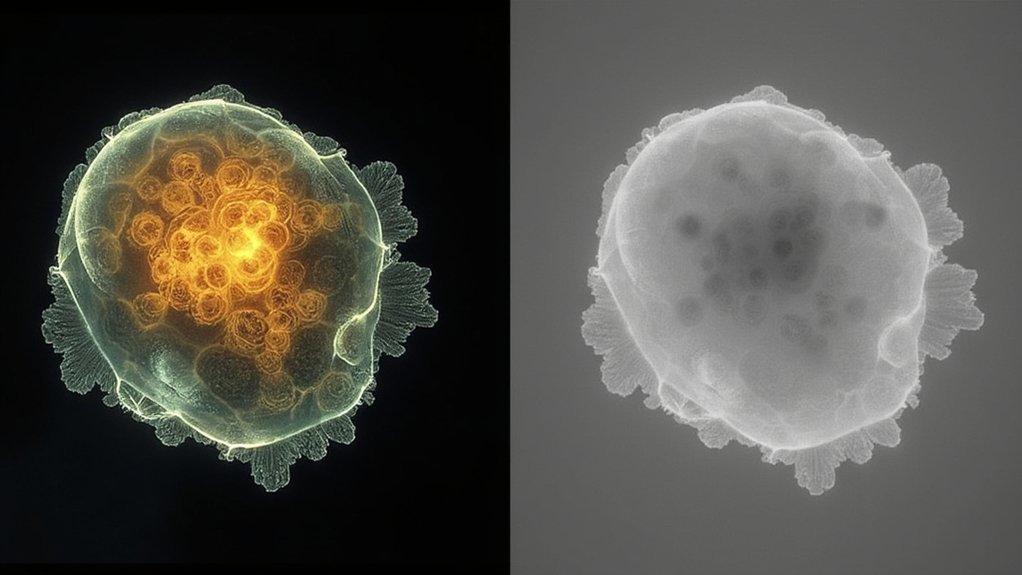
When selecting between dark field and phase contrast microscopy, understanding which specimens work best with each technique will greatly impact your results.
Dark field microscopy excels with live, unstained specimens like bacteria and small organisms, generating high contrast images against a dark background. You’ll find it ideal for highlighting minute details of delicate structures.
Phase contrast microscopy, on the other hand, is your go-to for transparent specimens, particularly live cells and their organelles. It reveals cell morphology without staining requirements and performs better with thicker specimens (5-10 micrometers).
While dark field can be combined with other techniques, phase contrast requires specialized optical components.
For biological research, consider your specimen type carefully—choose dark field for small, delicate organisms and phase contrast for observing cellular structures and behaviors in living tissue.
Equipment Setup and Technical Requirements
Setting up microscopy equipment for dark field and phase contrast imaging requires distinct optical configurations that directly impact image quality.
When preparing for dark field microscopy, you’ll need a specialized condenser with an opaque stop to create that distinctive bright image against a dark background.
Phase contrast microscopy, however, depends on precisely aligned phase rings in both condenser and objective lenses.
- Dark field requires angled illumination, while phase contrast needs a coherent light source (halogen or LED)
- You’ll need phase-specific objective lenses for phase contrast, but standard objectives work for dark field with the proper condenser
- Phase contrast systems demand more precise alignment of optical components
- Dark field setups are generally simpler, making them more accessible for beginners despite their specialized condenser requirements
Advantages and Limitations in Microscopic Photography
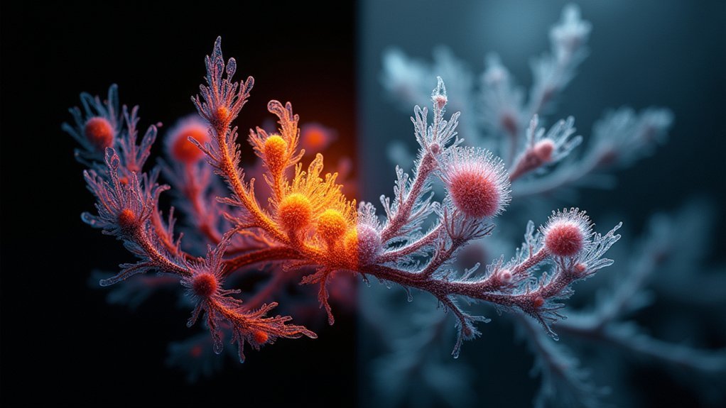
When you’re comparing microscopic photography techniques, you’ll find dark field excels at producing high-contrast images of unstained specimens while phase contrast converts optical variations into visible details without staining requirements.
Your specimen preparation differs considerably between methods, with dark field requiring minimal preparation for thin samples like bacteria, while phase contrast demands specialized optical components but handles a wider range of specimens.
You’ll achieve superior live cell visualization with both techniques, though dark field works better for observing motile organisms like Treponema pallidum, while phase contrast offers clearer views of internal structures and organelles in eukaryotic cells.
Contrast and Detail Capabilities
The fundamental distinction between dark field and phase contrast microscopy lies in their unique approaches to rendering invisible specimens visible. When capturing microscopic images, you’ll find dark field excels at highlighting small organisms against a black background, while phase contrast transforms transparent samples into high-contrast images without staining.
- Dark field microscopy creates striking contrast for unstained biological samples but may suffer from scattered light artifacts.
- Phase contrast microscopy reveals detailed internal cellular components by converting phase shifts into visible amplitude differences.
- Your specimen thickness considerably impacts results—phase contrast works best with specimens 5-10 micrometers thick.
- Each technique offers complementary visibility advantages—dark field enhances external structures while phase contrast reveals internal details.
Both methods have transformed microscopic photography, though each comes with specific limitations depending on your specimen characteristics and preparation methods.
Specimen Preparation Requirements
Specimen preparation stands at the heart of effective microscopic photography, with dark field and phase contrast offering significant advantages through their minimal preparation demands.
Both techniques allow you to capture live imaging of unstained specimens, eliminating time-consuming staining procedures that might alter cellular behavior.
With dark field microscopy, you’ll enjoy simpler setup requirements—perfect for thin samples where background noise can be minimized. You won’t need specialized optical components, making it more accessible and cost-effective.
Phase contrast microscopy requires more complex optical equipment but rewards you with high-contrast images revealing intricate cell morphology details.
Though slightly more demanding in setup, phase contrast excels when you’re studying internal cellular structures and behaviors in live cells, presenting clearer visualization of transparent features without the edge halos that sometimes obscure fine details.
Live Cell Visualization
Exploring live biological processes demands techniques that maintain cellular integrity while providing clear visualization, which is where dark field and phase contrast microscopy truly shine.
Both methods allow you to capture stunning photography of living specimens without staining, preserving their natural state.
- Dark field microscopy creates striking contrast against black backgrounds, making small organisms like bacteria pop visually while struggling with thicker specimens.
- Phase contrast microscopy reveals internal structures of transparent specimens by converting phase shifts to visible intensity differences.
- You’ll notice phase contrast excels with thin samples but produces halo artifacts that can obscure details in thicker specimens.
- When choosing between techniques, consider your specimen thickness and whether fine detail or overall structure visualization is your priority.
Advanced Techniques for Enhanced Image Quality
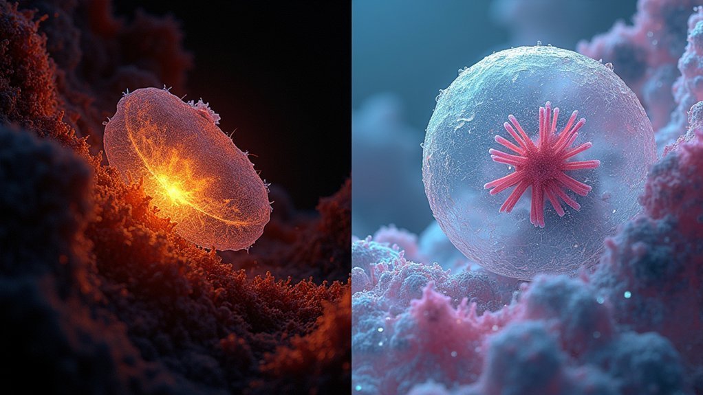
While both dark field and phase contrast microscopy offer significant advantages over traditional brightfield imaging, mastering advanced techniques can dramatically elevate your image quality.
When working with dark field microscopy, you’ll need to precisely align your specialized optics and adjust your condenser to create that striking contrast where unstained biological samples appear bright against a pitch-black background.
For transparent specimens, phase contrast microscopy requires proper positioning of phase rings to convert light wave shifts into visible amplitude differences, revealing internal details that would otherwise remain invisible.
Your choice between these methods should match your specific needs—use dark field when observing live microorganisms with external structures, and phase contrast when you need to visualize delicate internal components of cells.
Proper illumination control and aperture adjustment are essential for achieving enhanced contrast with either technique.
Frequently Asked Questions
What Is the Difference Between Dark Field and Phase Contrast?
Dark field creates bright images against dark backgrounds by capturing scattered light, while phase contrast transforms invisible phase shifts into visible contrast. You’ll find dark field simpler and cheaper, but phase contrast shows better internal details.
What Is the Difference Between Bright Field and Dark Field Imaging in TEM?
In bright field TEM, you’ll see a dark specimen against a bright background as electrons are absorbed. Dark field shows a bright specimen on a dark background, using only scattered electrons for better contrast.
What Are the Key Differences Between Brightfield and Phase-Contrast Microscopy?
You’ll notice brightfield microscopy shows stained cells as dark against a bright background, while phase-contrast transforms invisible phase differences into visible contrast, letting you observe living cells without staining them first.
What Is the Major Difference Between Phase Contrast and Differential Interference Contrast Microscopy?
The major difference is that phase contrast creates contrast by converting phase shifts into amplitude variations, while DIC uses polarized light and Nomarski prisms to create a 3D-like image with better boundary detail.
In Summary
You’ve now mastered the key differences between dark field and phase contrast microscopy. Each technique offers unique visual characteristics for your specimens, with dark field highlighting edges and phase contrast revealing internal structures. Choose dark field for dramatic contrast of transparent subjects or opt for phase contrast when you’re examining living cells. Whatever your microscopic photography needs, you’ll get better results by matching the technique to your specific application.


Leave a Reply