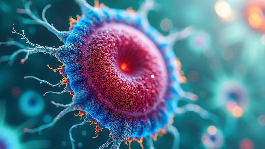
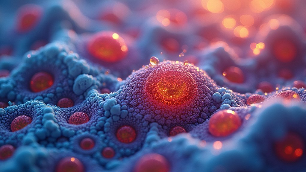
Staining helps you see specimen details better by creating visual contrast in otherwise transparent cells and microorganisms. Without stains, most biological structures have similar refractive indices, making boundaries nearly invisible under a microscope. Dyes bind to specific cellular components through chemical interactions, highlighting structures like cell walls, nuclei, and organelles. This differential coloration transforms invisible details into clearly distinguishable features, allowing you to identify pathogens and understand cellular architecture. The enhancement techniques reveal a microscopic world that would otherwise remain hidden.
The Optical Challenge of Unstained Specimens
When you first examine unstained specimens under a light microscope, you’ll immediately notice their frustrating lack of visual detail. This happens because cells and microorganisms are naturally transparent, allowing light to pass through with minimal scattering.
Without staining, you’ll struggle to distinguish essential cellular structures like cell walls and organelles from their surroundings due to poor contrast. The refractive index between different components is too similar, making boundaries almost invisible during optical microscopy.
This visibility problem becomes even more challenging when you’re trying to identify smaller, complex structures that require high-resolution imaging.
Staining addresses these limitations by enhancing the refractive index of specific components, reducing light scattering and creating the necessary contrast to transform those transparent, hard-to-see features into clearly defined, observable structures.
Chemical Mechanisms Behind Staining Techniques
Staining transforms invisible cellular details into vivid, observable structures through specific chemical interactions at the molecular level. When you apply dyes to specimens, their chromophores bind to specific cellular components based on chemical affinity.
Basic dyes carry positive charges that attach to negatively charged cell wall elements in a positive stain, while acidic dyes color the background in negative staining techniques.
Positively charged basic dyes bind to negative cell structures, while acidic dyes create contrast by coloring only the background.
The effectiveness of staining techniques often depends on chemical fixation with agents like formaldehyde, which preserves cellular morphology and enhances dye penetration.
In differential staining methods such as Gram staining, mordants like Gram’s iodine form insoluble complexes with primary dyes, improving retention. This chemical specificity allows you to distinguish bacterial types based on their cell wall composition, dramatically improving visibility and diagnostic capabilities in microscopy.
Differential Staining for Structure Identification

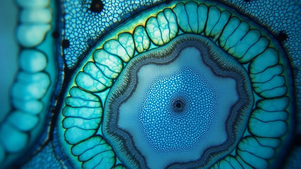
Although simple staining techniques offer basic visibility, differential staining methods provide remarkable structural clarity by employing multiple dyes that target specific cellular components. When you’re identifying bacteria, these techniques reveal essential details about cell wall composition that wouldn’t be visible otherwise.
- Gram staining uses crystal violet as a primary stain followed by Gram’s iodine and decolorization, turning gram-positive bacteria purple while gram-negative bacteria appear pink/red.
- Acid-fast staining enables visualization of mycobacteria with waxy cell walls, which retain carbolfuchsin and appear bright red against a blue background.
- Endospore and capsule staining use specialized approaches—heat-fixed malachite green penetrates resistant endospores, while negative staining creates contrast around capsules, revealing these cellular structures as clear halos.
Contrast Enhancement in Clinical Diagnostics
In clinical diagnostic laboratories, contrast enhancement through staining transforms cellular specimens from nearly invisible entities into clearly defined structures you can analyze with precision. Staining techniques dramatically improve visualization in clinical diagnostics by highlighting cellular structures that would otherwise remain indiscernible.
| Stain Type | Clinical Diagnostic Application |
|---|---|
| Gram | Identifies bacteria by cell wall composition |
| DAPI | Visualizes nuclei for cell type identification |
| Crystal Violet | Enhances contrast in tissue biopsies |
| Methylene Blue | Reveals cellular morphology changes |
| Fluorescent Dyes | Enables real-time observation of living cells |
You’ll find that differential techniques like Gram staining provide critical information for pathogen identification, while fluorescent dyes allow you to monitor cellular processes in real time, substantially improving your ability to detect abnormalities and make accurate diagnoses.
Modern Digital Enhancement of Stained Specimens
While traditional staining methods revolutionized microscopy, digital enhancement technologies have greatly transformed how you interpret stained specimens. High-resolution digital microscopy coupled with sophisticated image processing algorithms substantially improves visibility by reducing noise and enhancing contrast in your stained preparations.
- Virtual staining techniques allow you to apply different color maps digitally, offering flexibility in visualizing cellular structures without additional physical stains.
- Automated image analysis tools quantify stain intensity and distribution, helping you identify cellular abnormalities with greater diagnostic accuracy.
- Integration of fluorescence microscopy with digital imaging enables real-time observation of dynamic biological processes in live cells.
These digital enhancements don’t replace traditional staining but complement it, giving you unprecedented clarity and analytical power when examining microscopic specimens.
Frequently Asked Questions
What Is the Advantage of Staining Specimens?
Staining gives you increased visibility of cellular structures by enhancing contrast. You’ll see transparent specimens clearly, identify specific components like nuclei and cell walls, improve resolution, differentiate cell types, and detect abnormalities more effectively.
How Was the Stain Helpful in Observing the Specimen?
The stain helped you observe the specimen by enhancing contrast, making transparent structures visible, highlighting specific cellular components, and differentiating between cell types that wouldn’t be distinguishable under normal microscopic examination.
Why Is Stain Needed to Better See the Cells?
Stains are needed because they enhance contrast, allowing you to see otherwise transparent cells. They’ll bind to specific cellular components, highlighting structures and making it easier for you to identify different cell types.
What Is the Purpose of Using a Stain When Viewing a Tissue Sample?
You’ll use stains to enhance visibility of transparent tissue samples, highlight specific cellular structures, reveal metabolic processes, differentiate between live and dead cells, and preserve tissue morphology for detailed microscopic analysis.
In Summary
You’ve seen how staining transforms invisible cellular details into vibrant, distinguishable structures. By selectively binding to specific components, stains create the contrast your eyes need to differentiate otherwise transparent features. Whether you’re examining bacteria, tissues, or cellular organelles, staining techniques remain essential tools that bridge the gap between microscopic structures and your ability to visualize them in meaningful, diagnostic ways.


Leave a Reply