Category: Staining Techniques in Microscope Photography
-
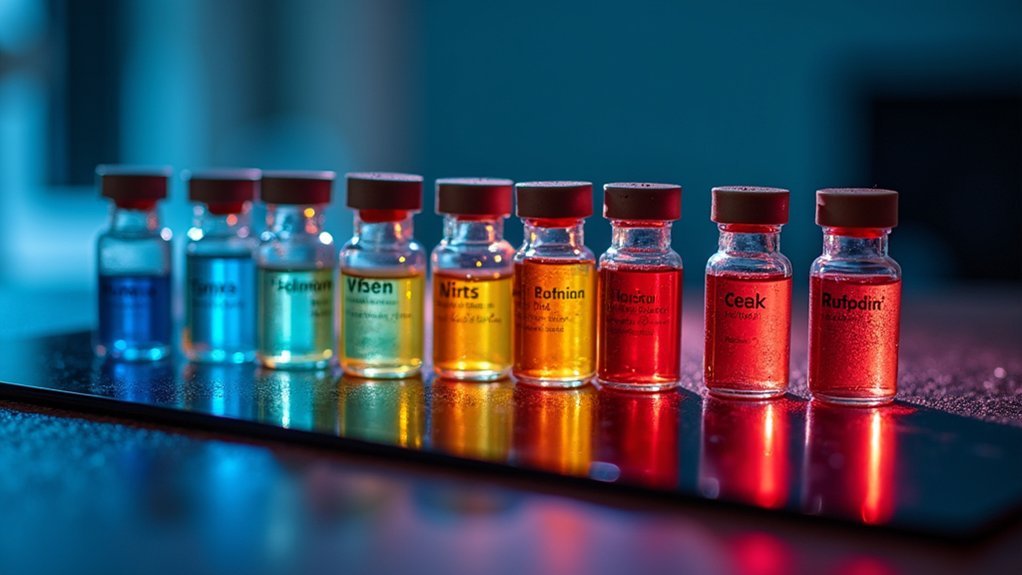
7 Best Vital Dyes for Living Cell Observation
Astonishing vital dyes revolutionize live cell imaging, revealing dynamic cellular processes while maintaining viability—but which seven outperform all others?
-
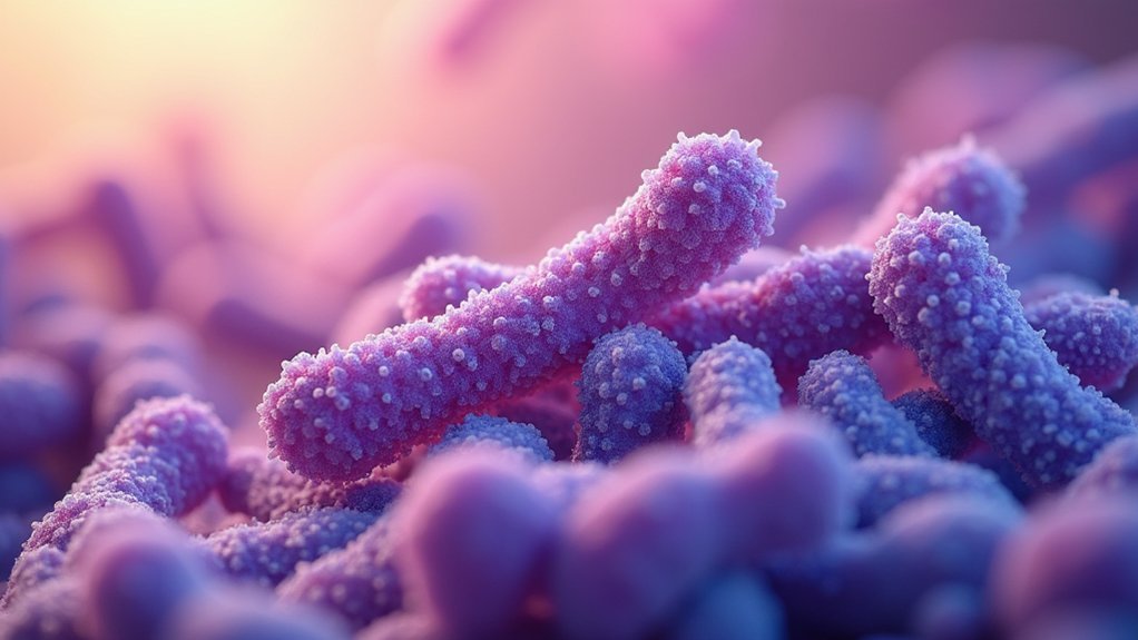
7 Tips For Stunning Gram-Stained Bacteria Photos
Achieve jaw-dropping bacterial photography with these essential gram staining techniques that transform ordinary specimens into mesmerizing microscopic art.
-
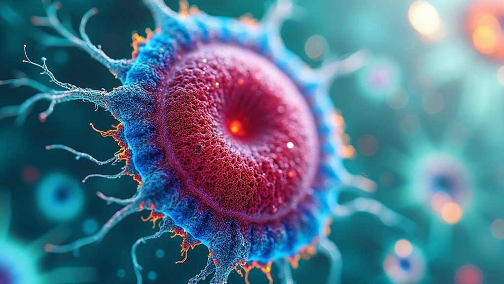
3 Best Methods For Capturing Fluorescent Specimen Images
Achieving stunning fluorescent specimen images requires optimized fixation, compatible fluorophores, and fine-tuned microscope settings—but which technique matters most?
-
Why Does Staining Help See Specimen Details Better?
From nearly invisible to vividly detailed, staining transforms microscopic specimens by creating contrast that reveals hidden cellular structures.
-
Expert Guide: Differential Staining for Clear Specimen Images
Behind every striking bacterial image lies essential staining techniques that transform invisible microbes into vivid, diagnostic specimens.
-
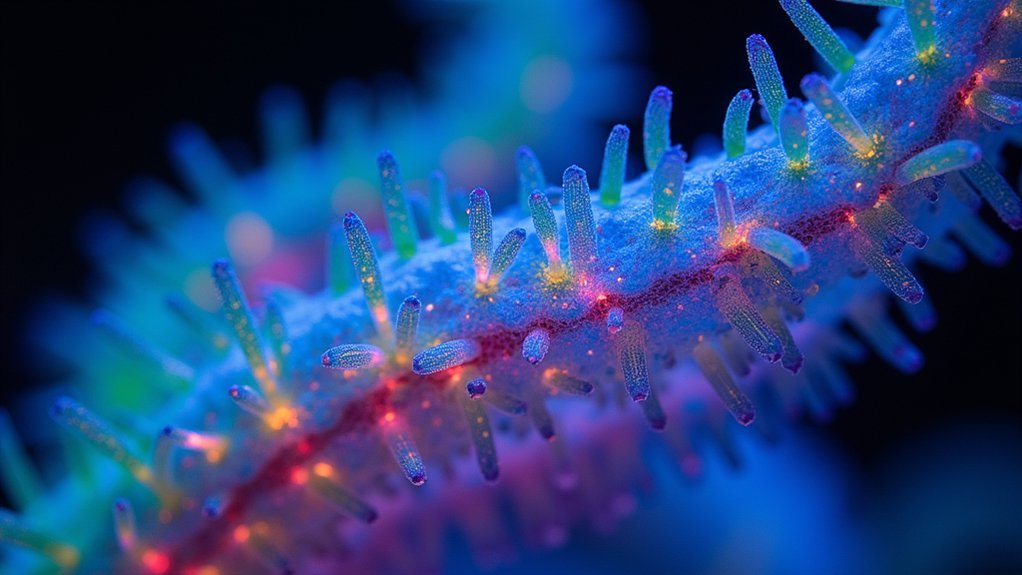
3 Best Negative Stains for Revealing Cell Structure
Featuring uranyl acetate, PTA, and ammonium molybdate, these negative stains reveal cellular mysteries invisible to the naked eye.
-

Top Stains For Crystal-Clear Specimen Photography
Outstanding microscopy stains transform ordinary specimens into vibrant visual narratives, but which formulation delivers the most photogenic results?