For ideal dark field bacterial photography, center your condenser and verify its numerical aperture exceeds your objective’s. Prepare bacteria on thin glass slides (1.1-1.2mm) to maximize contrast. Use high-intensity LED lighting with controlled oblique illumination. Apply short exposure times (1/60-1/1000s) with appropriate ISO settings to balance detail and noise. Enhance your images post-capture by adjusting contrast, reducing noise, and applying selective sharpening. These techniques will transform invisible bacteria into brilliantly illuminated specimens against a black backdrop.
Condenser Alignment and Numerical Aperture Optimization
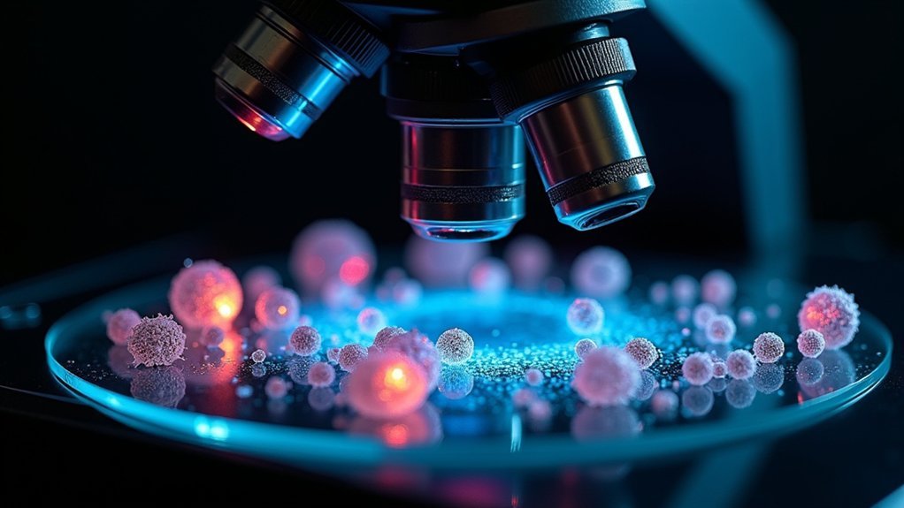
When capturing bacterial images in dark-field microscopy, proper condenser alignment serves as the foundation for exceptional results. You’ll need to center your condenser precisely to prevent uneven illumination and maximize the visibility of refracted light from bacterial specimens.
Any misalignment introduces glare and diminishes image clarity due to dust particle interference.
Always verify your dark-field condenser’s numerical aperture exceeds that of your objective lens. This critical relationship enables effective light collection around your specimen.
For high magnification objectives (60X and 100X), use specialized condensers with an iris diaphragm to control illumination conditions.
Don’t overlook your slide preparation—clean glass slides between 1.1 and 1.2 mm thick are essential.
Thicker slides scatter light inappropriately and obscure crucial details in your bacterial specimens, undermining your efforts to achieve striking dark-field photography.
Specimen Preparation for Maximum Contrast
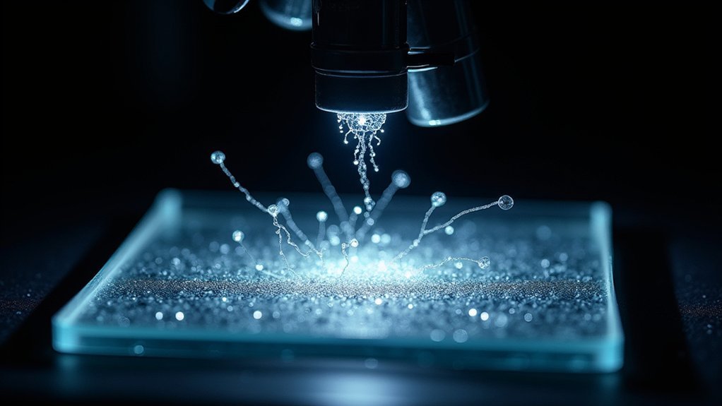
Effective specimen preparation forms the cornerstone of high-contrast dark-field bacterial photography. You’ll need clean glass slides that are 1.1-1.2mm thick to guarantee ideal light transmission through your specimens.
Always prepare bacteria in thin sections to minimize diffraction artifacts, which greatly enhances fine structural details under dark-field illumination.
Thin bacterial preparations reduce diffraction, revealing intricate structural details that might otherwise remain hidden under dark-field conditions.
Your condenser and numerical aperture settings must be perfectly adjusted to maximize contrast in light microscopy. Use high-intensity oblique light sources to illuminate your specimens effectively, as dark-field illumination relies on this angular lighting to create striking contrast against a black background.
Don’t overlook regular cleaning of all optical surfaces. Even minor smudges or dust can scatter light improperly, diminishing the quality of your high-contrast images.
Meticulous preparation of specimen slides ultimately determines the clarity and scientific value of your bacterial photographs.
Controlling Light Source Intensity and Direction
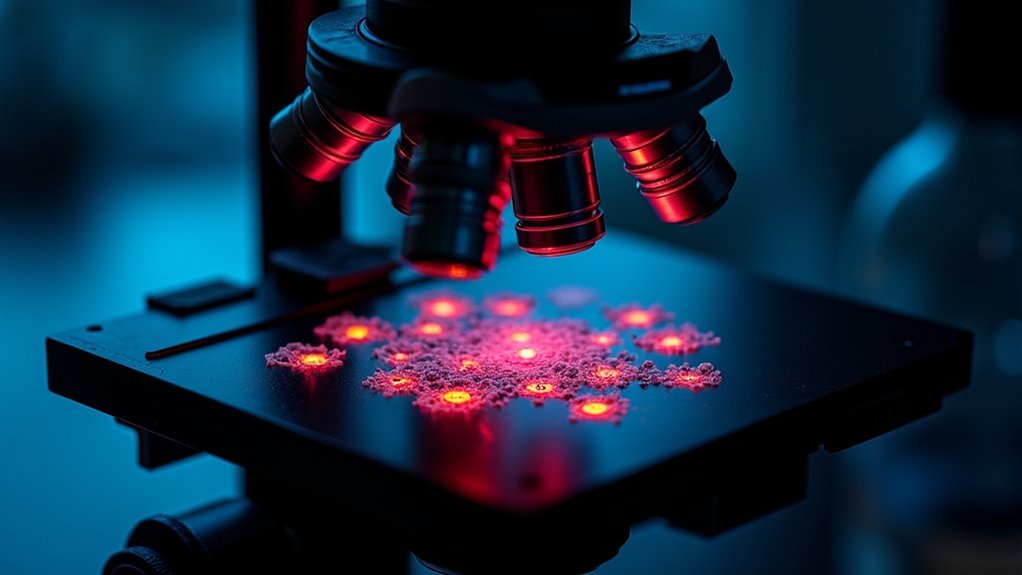
Three critical factors determine the success of dark-field bacterial photography: light source intensity, direction, and control.
You’ll need a high-intensity LED or fiber optic lamp to compensate for the limited light that reaches your camera sensor.
Position your light source to achieve oblique illumination, creating contrast between bacteria and the dark background by capturing only scattered light through the objective lens.
Your dark-field condenser’s iris diaphragm is essential for controlling numerical aperture and light intensity.
For ideal results:
- Clean all optical components regularly to eliminate dust that creates unwanted glare
- Adjust your light source angle until bacteria appear bright against the background
- Experiment with various exposure times and gain settings to balance detail capture while minimizing noise
Maintaining precise control over these elements transforms ordinary bacterial specimens into striking scientific images.
Camera Settings for Optimal Bacterial Visualization
Selecting the right camera settings directly impacts your success in dark-field bacterial photography, as even the most perfectly illuminated specimen can appear washed out or grainy with improper configuration.
Use short exposure times (1/60 to 1/1000 seconds) to prevent overexposure from intense light sources typical in dark-field microscopy.
Rapid exposures capture bacterial brilliance without washing out delicate structures in dark-field imaging.
Increase camera gain to enhance sensitivity in low-light situations, but find the balance that minimizes noise.
Opt for lower ISO settings (100-200) to reduce noise when capturing bright bacterial specimens against dark backgrounds.
Experiment with aperture settings to control depth of field—wider apertures can beautifully isolate your bacterial specimens by blurring the background.
Don’t forget to use histogram analysis to guarantee proper dynamic range, avoiding clipped highlights or shadows for the clearest visualization of bacterial structures and movement.
Post-Processing Techniques for Enhanced Image Clarity
Once you’ve captured your bacterial specimens through dark-field microscopy, thoughtful post-processing transforms good images into exceptional ones with greater scientific value.
Adjusting brightness and contrast emphasizes the distinction between bright bacterial structures and the dark background, revealing details that might otherwise remain hidden.
For ideal image clarity in your dark field images:
- Apply noise reduction tools to eliminate bright spots from dust or contaminants that can distract from bacterial details.
- Utilize sharpening filters to enhance cell edges, making bacterial structures more defined against the dark background.
- Consider strategic cropping and resizing to focus on your area of interest while removing distracting elements.
Color adjustments can further improve both aesthetic appeal and scientific value, ensuring important bacterial features remain visible and interpretable for analysis.
Frequently Asked Questions
Which Is the Most Effective Use of Dark Field Microscopy?
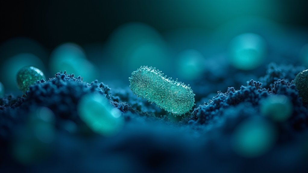
Dark field microscopy’s most effective use is visualizing live, unstained bacteria. You’ll see transparent specimens clearly against a dark background, revealing fine structures like flagella that you’d miss with traditional microscopy techniques.
What Is the Darkfield Technique?
Darkfield technique is a microscopy method where you’ll see specimens illuminated against a dark background. You’re using oblique light that scatters when hitting your sample, making transparent organisms like bacteria strikingly visible without staining.
How to Prepare a Slide of an Ideal Specimen for a Dark Field Microscope?
To prepare an ideal slide for dark field microscopy, you’ll need a thin specimen (under 1mm), clean optical surfaces, transparent or lightly pigmented material, matching refractive index mounting medium, and proper condenser alignment.
What Is the Dark Field Microscopy for Bacteria?
Dark-field microscopy lets you observe bacteria as bright images against a dark background. You’ll see unstained living bacteria clearly, including tiny structures like flagella that you couldn’t view with standard microscopy techniques.
In Summary
Mastering dark field bacterial photography isn’t as intimidating as you might think. By properly aligning your condenser, preparing specimens carefully, controlling your light source, optimizing camera settings, and applying targeted post-processing techniques, you’ll capture stunning bacterial images that reveal incredible detail. With practice, you’ll develop your own workflow that consistently produces clear, high-contrast photographs worthy of both scientific documentation and artistic appreciation.


Leave a Reply