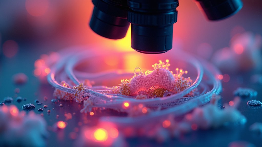
For stunning brightfield microscope photos, you’ll want to master Köhler illumination for even light distribution. Set your condenser aperture to one-third of the objective’s numerical aperture, adjust color temperature to 5500K, and use ISO 100-200 with 1/15 second shutter speed. Properly prepare specimens with appropriate stains, employ focus stacking for depth, and save images in lossless formats. These seven adjustments will transform your ordinary microscope views into publication-worthy scientific imagery.
Mastering Köhler Illumination for Even Light Distribution

The foundation of any quality brightfield microscope image begins with proper illumination. Köhler illumination stands as the gold standard for achieving even illumination across your entire field of view, eliminating shadows and bright spots that compromise image quality.
Perfect illumination forms the foundation of microscopy excellence, with Köhler technique eliminating imperfections that mask critical specimen details.
To establish proper Köhler illumination, start by fully opening both your condenser and field iris diaphragms. After focusing on your specimen, partially close the field iris until its edges appear sharp in your view.
Then verify your condenser is perfectly aligned with your objective—any misalignment will immediately degrade your results.
This technique markedly enhances contrast in brightfield microscopy, revealing fine details that would otherwise remain hidden.
For stunning photomicrographs, regularly calibrate your setup to maintain ideal performance. Your extra effort in mastering Köhler illumination will be immediately visible in your images.
Optimizing Condenser Aperture Settings for Depth and Contrast
To achieve ideal image quality, you’ll want to position your condenser aperture at roughly one-third of your objective’s numerical aperture, creating the perfect balance between resolution and contrast.
When searching for your specimen’s contrast sweet spot, try adjusting the iris diaphragm incrementally and notice how different settings affect visibility and depth perception.
Don’t forget to properly align your field diaphragm as well, ensuring it’s centered and adjusted appropriately to complement your aperture settings for crisp, evenly illuminated observations.
Proper Aperture Positioning
Why does your brightfield microscope image look flat and washed out? The answer likely lies in your condenser aperture positioning. For ideal results, you’ll want to adjust the aperture to allow only one-third to one-half of the objective aperture to remain open. This critical balance enhances contrast while maintaining clarity without introducing excessive graininess.
Proper positioning isn’t just about light intensity—it greatly improves your depth of field, allowing you to focus clearly on different specimen layers. Remember that different objectives require specific settings; lower magnifications benefit from wider apertures, while higher powers need more restricted openings.
For the best resolution, match your condenser iris to the numerical aperture of your objective lens. This creates a focused, uniform light beam that illuminates your specimen perfectly. Regular calibration prevents uneven illumination and unwanted artifacts in your photographs.
Finding Contrast Sweet Spot
Achieving perfect contrast in brightfield microscopy requires finding the delicate balance between too much and too little light.
Begin with your condenser aperture fully open while focusing, then gradually close it to approximately one-third of your objective’s numerical aperture—this is your contrast sweet spot.
Watch how the illumination changes as you adjust; a smaller aperture increases depth of field but may reduce brightness. You’ll know you’ve found the ideal setting when specimen details appear crisp without sacrificing clarity or introducing diffraction artifacts.
Proper Köhler illumination techniques guarantee even light distribution across your field of view, enhancing your ability to fine-tune contrast.
Remember to regularly check your optical components for alignment and cleanliness, as even minor issues can greatly diminish image quality, no matter how perfect your aperture settings.
Field Diaphragm Adjustments
While the condenser aperture controls contrast, your field diaphragm serves as the gatekeeper for incoming light, dramatically influencing image quality. Properly adjusting this component is vital for successful photomicrography with brightfield microscopes.
The ideal setting requires a delicate balance between illumination and contrast. Start by partially closing the field diaphragm from full open to enhance detail without causing vignetting. Verify proper alignment with the condenser aperture for even illumination across your specimen.
- Close slightly – Enhance depth of field and reveal fine structures
- Find the balance – Too open causes glare, too closed darkens periphery
- Adjust per specimen – Different samples demand unique settings
- Check alignment – Proper centering guarantees even illumination across the field
Different specimens require different field diaphragm positions, so experiment to find each sample’s sweet spot.
Perfect Color Temperature Adjustments for True-to-Life Imaging
How often have you captured a microscopic image only to find the colors don’t match what you observed through the eyepiece? Achieving accurate color representation starts with adjusting your microscope’s color temperature to approximately 5500K for daylight-balanced illumination.
Color fidelity in microscopy begins with proper illumination—set your microscope’s color temperature to 5500K for truthful specimen representation.
Don’t overlook your camera’s white balance—set it to “daylight” or “manual” to prevent unwanted color casts.
For ideal color accuracy, use color correction filters to match your light source with your camera’s sensor settings. Include a color chart during setup to calibrate and make necessary adjustments based on observed discrepancies.
Remember that dirty optical components can distort colors, so clean them regularly to maintain light quality.
These simple adjustments will guarantee your microscope images faithfully reproduce the vibrant details and true colors of your specimens.
Strategic Camera Exposure Settings for Brightfield Clarity

To capture the intricate details visible through your brightfield microscope, you’ll need to carefully configure your camera’s exposure settings.
Digital cameras require specific adjustments to produce high contrast, crisp images that truly represent your specimen.
- Set your shutter speed to approximately 1/15 second – this sweet spot allows adequate light collection while preventing motion blur for stationary specimens.
- Keep your ISO setting low (100-200) to minimize grain and preserve fine details in brightfield conditions.
- Adjust your white balance appropriately for your light source – automatic works well for most setups, but manual tweaking can dramatically improve color accuracy.
- Open your condenser iris to no more than one-third of the objective aperture – this prevents graininess while maintaining brightness and ideal detail resolution.
Fine-Tuning Focus Stacking Techniques for Extended Depth of Field
When examining specimens at high magnifications, you’ll quickly notice the limited depth of field restricts your view to a narrow focal plane.
Focus stacking solves this problem by combining multiple images captured at different focal points, especially vital at 20X and above on your light microscope.
Using your digital SLR, capture 3-40 images at sequential focus levels while maintaining consistent lighting and camera settings.
Proper sample preparation is essential before beginning this process to guarantee stability throughout the sequence.
Import your images into specialized software like Helicon Focus or Photoshop, which will align and merge them into a composite with dramatically extended depth of field.
This technique reveals details that would otherwise remain partially blurred, resulting in good photomicrographs that accurately represent your specimen’s three-dimensional structure.
Specimen Preparation Methods to Enhance Visual Detail

Exceptional specimen preparation forms the foundation of all high-quality brightfield microscopy. Your specimens must be properly mounted on clean glass slides with appropriate coverslips to eliminate artifacts and maximize light transmission.
When staining specimens, select reagents compatible with your sample type to highlight specific cellular structures and create necessary contrast.
For stunning brightfield photos, remember to:
- Keep specimens hydrated throughout observation to maintain structural integrity
- Section specimens to 5-10 micrometers thickness for peak light penetration
- Apply anti-fade reagents when using fluorescence to prevent photobleaching
- Use coverslips that match your objective’s specifications for aberration-free imaging
These preparation techniques will dramatically enhance detail visibility and produce images with greater clarity, color fidelity, and scientific value than poorly prepared specimens ever could.
Digital Processing Workflows for Professional Microscope Images
Professional microscope images require three essential digital processing steps to transform raw captures into publication-quality visuals.
First, calibrate your imaging system to maintain consistent light source settings across experiments, ensuring reliable comparison between specimens.
Next, enhance your microscopy images using software like Photoshop or ImageJ to optimize contrast and brightness while preserving specimen integrity.
Apply histogram equalization techniques to improve dynamic range, distributing pixel intensities evenly without losing detail in highlights or shadows.
Finally, save your processed images in lossless formats like TIFF or PNG to prevent data degradation.
Consider focus stacking for specimens requiring greater depth of field—this technique combines multiple focal plane images into one sharper composite, revealing intricate details that single captures might miss.
Frequently Asked Questions
What Camera Settings Are Needed for Microscope Photography?
For microscope photography, you’ll need 1/15 second shutter speed, ISO 100-200, automatic white balance, and manual focus. Consider using an electronic shutter release to minimize vibration for sharper, clearer specimen images.
How to Enhance Microscope Images?
You’ll enhance microscope images by using Köhler illumination, selecting appropriate contrast techniques, keeping optics clean, optimizing camera settings with low ISO, and investing in quality objective lenses like Plan Achromat types.
How Do You Adjust a Microscope for Optimal Viewing?
For ideal microscope viewing, you’ll need to establish Köhler illumination, adjust the condenser height, partially close the field iris, and use Plan Achromat objectives. Don’t forget to clean all optics regularly.
How Do You Take Good Pictures With a Microscope?
To take good pictures with a microscope, you’ll need proper Köhler illumination, clean optics, and stable camera settings. Use a low ISO, moderate shutter speed, and experiment with focus stacking for sharper high-magnification images.
In Summary
You’ll achieve stunning brightfield microscopy images by combining these seven essential settings. Mastering Köhler illumination, optimizing aperture settings, and adjusting color temperature form your foundation. When you complement these with proper exposure, focus stacking, and careful specimen preparation, your images will stand out. Don’t overlook the final digital processing step—it’s what transforms good microscope photos into exceptional scientific visuals that truly communicate your findings.


Leave a Reply