
For crystal-clear specimen photography, you’ll achieve best results with Safranin (red) for cellular structures, Methylene Blue for nuclear definition, and Eosin Y for vascular highlights in botanical samples. Janus Green specifically targets chloroplasts with vibrant blue differentiation. Consider combining complementary stains like Safranin with Light Green for multi-dimensional imaging. Aceto Carmine creates dramatic contrast for inversion techniques. Pair these with polarizing filters to transform ordinary specimens into compelling visual narratives.
Safranin: The Cellular Structure Revealer
A powerhouse in microscopic visualization, Safranin transforms ordinary specimen photographs into revelatory scientific images. You’ll find this vibrant red stain particularly valuable when you need quite accurate differentiation between lignified and non-lignified cells in plant tissues.
When photographing specimens like Azalea stems, immerse segments in Safranin for several minutes, ensuring proper cellular penetration. This technique highlights otherwise obscured details, making your microphotography more informative and visually striking.
For enhanced results, combine Safranin with complementary stains to create contrast that showcases nuclei and vascular structures. This approach is required for thorough tissue analysis, revealing intricate organizational patterns within your samples.
The stain’s ability to emphasize cellular architecture makes it indispensable for both educational demonstrations and research documentation.
Methylene Blue for Enhanced Cellular Definition
When detail matters most in your specimen photography, Methylene Blue stands out as an exceptional stain that transforms ordinary samples into vivid displays of cellular architecture. This crucial stain provides strong contrast by imparting a distinct blue color that highlights nuclei and organelles in both plant and animal specimens.
You’ll find Methylene Blue particularly effective when working with plant tissues like Azalea stems, where it reveals intricate cellular arrangements with remarkable clarity.
The staining process is invigoratingly quick—you’ll only need a few minutes of exposure to achieve ideal results without overwhelming your specimen. After application, simply wash away excess stain with purified water to prevent distortion and enhance image clarity during microphotography.
Achieve vibrant definition in minutes, then rinse with purified water for crystal-clear microphotography results.
It’s this balance of vivid definition and ease of use that makes Methylene Blue essential for your microscopy toolkit.
Eosin Y: Natural Enhancement for Botanical Specimens
While Methylene Blue excels at revealing cellular architecture, Eosin Y offers photographers a complementary approach with its distinctive orange-pink coloration. You’ll find this synthetic dye particularly valuable when photographing plant specimens, especially when highlighting vascular structures like protoxylem vessels in carrots.
| Specimen Type | Benefits of Eosin Y | Best Application |
|---|---|---|
| Carrot tissue | Highlights protoxylem | After sectioning |
| Stem sections | Reveals vascular bundles | Wash excess with water |
| Leaf samples | Enhances cellular boundaries | Use with microscope |
| Root tissues | Improves structural contrast | Keep specimen moist |
| Floral parts | Defines tissue layers | Combine with other stains |
After staining, rinse with purified water to maintain moisture and definition. When capturing microphotographs, you’ll notice the enhanced visibility of cellular arrangements, making Eosin Y indispensable for your botanical imaging toolkit.
Janus Green: Highlighting Chloroplasts and Epidermal Cells
Janus Green stands out as a specialized essential stain that transforms your plant specimen photography by creating striking blue differentiation in microscopic images. This powerful stain specifically targets chloroplasts, the photosynthetic powerhouses of plant cells, making them clearly visible against surrounding cellular structures.
You’ll achieve high-contrast microphotography by applying the Janus Green solution briefly to your specimen, then rinsing to eliminate excess stain. This prevents unwanted background coloration while preserving the vivid blue highlights on your target structures.
The resulting images reveal intricate details of both chloroplasts and epidermal cells with remarkable clarity.
For researchers and photographers documenting plant cellular architecture, Janus Green provides the visual distinction needed to analyze tissue composition effectively, making it an indispensable tool in your botanical imaging arsenal.
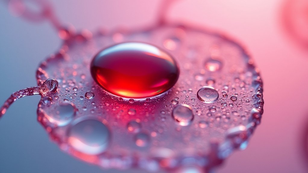
Aceto Carmine Staining for Dramatic Contrast
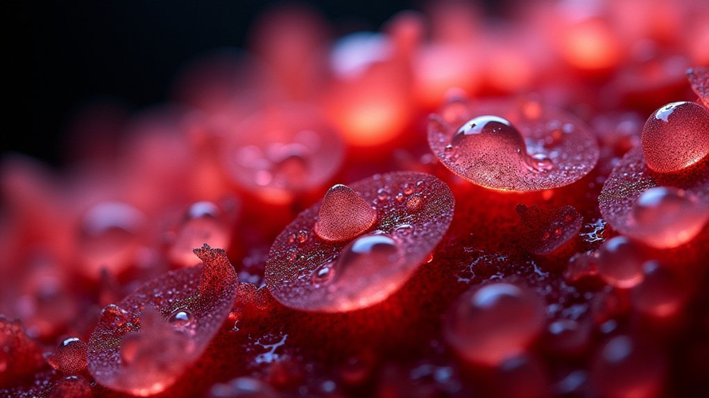
Aceto Carmine’s powerful red coloration reveals cellular structures with exceptional clarity, transforming ordinary plant specimens into visually striking images.
You’ll find this stain particularly effective when employing inversion color techniques in your post-processing workflow, as the dramatic red-against-background contrast creates compelling depth.
Cellular Structure Revelation
Three key qualities make Aceto Carmine staining indispensable for specimen photography: its powerful nucleic acid binding properties, vivid red coloration, and exceptional contrast enhancement.
When you’re trying to capture intricate cellular arrangements, this stain reveals details that remain invisible to the naked eye.
For best results, you’ll need to allow sufficient staining time—typically several minutes—ensuring the dye thoroughly penetrates your plant tissue specimens.
Keep your preparations consistently moist during the imaging process; this prevents fading and maintains the vibrant visualization of cellular structures.
You’ll find Aceto Carmine particularly effective for distinguishing between different cell types in plant tissues.
The dramatic red-against-background contrast it creates makes your microphotography pop with clarity, highlighting structural details that might otherwise go unnoticed.
Inversion Color Techniques
Employing advanced inversion color techniques with Aceto Carmine staining allows you to transform ordinary specimen photographs into dramatic visual revelations.
This powerful red stain creates exceptional contrast that highlights cellular details in plant tissues that would otherwise remain invisible to your camera lens.
When working with Aceto Carmine, remember these essential steps:
- Monitor your staining time carefully to prevent oversaturation that might obscure delicate cellular structures.
- Apply the inversion color technique to create striking visual effects that emphasize specific tissue components.
- Fine-tune the inverted images using RGB Levels in Capture One Pro to achieve both scientifically accurate and artistically compelling results.
You’ll find this method particularly effective for plant specimens, where the vivid contrast reveals intricate cellular architecture with unprecedented clarity.
Visual Effect Enhancement
To achieve truly remarkable specimen photographs, mastering visual effect enhancement with Aceto Carmine staining is essential. This powerful water-soluble stain binds to nucleic acids, creating vivid red coloration against lighter backgrounds that dramatically highlights cellular structures.
You’ll find Aceto Carmine particularly valuable when photographing plant tissues during cell division, as it makes chromosomes stand out with striking clarity. The stain’s water solubility allows you to apply it easily and rinse excess away without damaging delicate specimens.
For scientific documentation requiring precise cellular detail, combine Aceto Carmine with appropriate lighting techniques. You’ll capture images where cell walls and nuclei are clearly delineated with impressive contrast.
This combination of staining and proper illumination transforms ordinary microscopy into compelling visual documentation that reveals the intricate architecture of your specimens.
Light Green Stain for Vascular Bundle Visualization
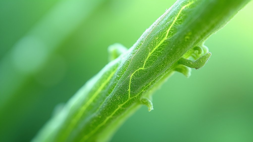
When examining plant specimens under the microscope, light green stain stands out as an excellent choice for visualizing vascular bundles. This specialized dye enhances the transparency of thin sections, allowing light to penetrate and highlight the intricate network of xylem and phloem within stems and leaves.
To achieve ideal results with light green stain, follow these simple steps:
- Apply the stain to your prepared plant section and allow it to work for just a few minutes.
- Wash thoroughly with purified water to remove excess dye that might cloud your image.
- Mount and photograph immediately to capture the enhanced contrast between vascular tissues.
You’ll find this technique particularly valuable when you’re studying the arrangement and physiological functions of plant transport systems in your microphotography work.
Combining Stains for Multi-Dimensional Imaging
You’ll achieve striking visual depth by layering complementary dyes like Safranin with Janus Green to reveal multiple cellular structures simultaneously.
Test your stain combinations on sample sections first to verify they enhance rather than mask each other’s visibility.
For maximum contrast enhancement, try pairing Eosin Y with Light Green stain to highlight both protoxylem vessels and vascular bundles in your specimen photographs.
Layering Complementary Dyes
While single stains provide valuable visualization, the art of layering complementary dyes transforms specimen photography into a multi-dimensional experience. By combining stains like Eosin Y and Methylene Blue, you’ll reveal intricate cellular structures that would otherwise remain hidden, creating images with enhanced contrast and detail.
For successful layering results:
- Test compatibility – Not all dyes play well together. Verify that your chosen stains won’t react negatively when combined, preserving image clarity.
- Time your application – Different staining durations can dramatically affect visualization. Experiment with timing to achieve ideal differentiation of cellular components.
- Focus on complementary targets – Select dyes that highlight different structures, such as one for chloroplasts and another for vascular bundles, to maximize the informational content of your microphotographs.
Contrast Enhancement Techniques
The magic of contrast enhancement in specimen photography lies in strategic stain combinations.
When you pair complementary stains like Safranin for vascular tissue with Janus Green for chloroplasts, you’ll create striking visual distinctions between cellular structures within the same specimen.
You can fine-tune your results by adjusting staining duration for each dye, optimizing color retention where it matters most.
Try layering Eosin Y over protoxylem vessels before applying a secondary stain to add depth and enhance three-dimensional qualities in your final images.
Don’t hesitate to experiment with application order and dilution ratios.
These variables often reveal intricate details invisible with single-stain approaches.
The most compelling specimen photographs emerge when you’ve thoughtfully combined stains to highlight specific cellular features while maintaining overall visual clarity.
Polarizing Filters With Stains for Artistic Enhancement
Beyond their scientific utility, polarizing filters transform stained specimen photography into a fascinating art form.
You’ll notice immediately how they reduce glare while dramatically enhancing contrast when working with stains like Eosin Y or Janus Green.
- Experiment with filter positioning relative to your light source—this simple adjustment creates stunning artistic effects that showcase how light interacts with your specimens.
- For plant materials such as Azalea or cucumber stems, polarizing filters reveal intricate vascular bundles and chloroplasts with vivid clarity you can’t achieve otherwise.
- Combine these filters with high-resolution imaging techniques to create visual documentation that’s both scientifically valuable and aesthetically striking.
This creative approach doesn’t just improve visibility—it transforms your microphotography into compelling visual narratives that highlight the hidden beauty of cellular structures.
Specimen-Specific Stain Selection Guide
Selecting appropriate stains for your specimens can dramatically transform ordinary microscopy images into revealing scientific documents.
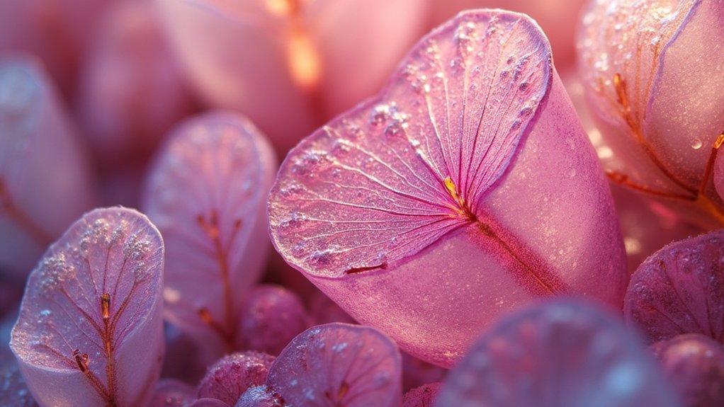
For Azalea stem sections, choose Safranin stain to achieve vibrant red highlights that accentuate cellular structures.
When working with carrot samples, Eosin Y delivers ideal orange-pink coloration that beautifully reveals protoxylem vessels and their intricate details.
If you’re photographing specimens with chloroplasts or need to emphasize epidermal cells, try Janus Green stain, which appears more blue than green despite its name.
For cucumber stems, Light Green stain will make vascular bundles pop under the microscope, enhancing your analytical capabilities.
Remember that both your choice of stain and staining duration considerably impact the final image quality.
Always select your stain based on the specific specimen and the structures you’re aiming to highlight.
Frequently Asked Questions
What Is the Best Way to Photograph Crystal?
Use a diffuse light source, tripod for stability, and macro lens to capture intricate details. You’ll want to experiment with different angles and backgrounds. Consider polarized filters to reduce reflections and enhance crystal clarity.
Why Are Stains Often Used in Microscopy and What Is the Benefit of Using Different Stains Even on the Same Sample )?
Stains in microscopy improve contrast, making structures visible. You’ll benefit from using different stains on the same sample as they highlight various cellular components – revealing cell walls, organelles, and other features you’d otherwise miss.
In Summary
You’ll achieve spectacular photomicrographs by selecting the right stain for your specimen type. Don’t hesitate to experiment with combinations that enhance specific structures. Remember, proper dilution and application techniques are as important as the stain itself. With these professional-grade stains and methods, you’re now equipped to capture crystal-clear images that reveal the microscopic world in stunning detail.


Leave a Reply