Fluorescent stains create the most visually spectacular biological images by transforming invisible cellular structures into vibrant light shows. You’ll find immunofluorescence particularly striking, with its multicolored protein targeting capabilities. Gram staining offers classic blue-purple and pink contrasts, while Romanowsky stains transform blood smears into detailed, colorful maps of different cell types. DIC microscopy reveals living specimens’ natural beauty without staining at all. The intersection of science and art continues beyond these remarkable visualization techniques.
Fluorescent Stains: Creating Vibrant Cellular Landscapes
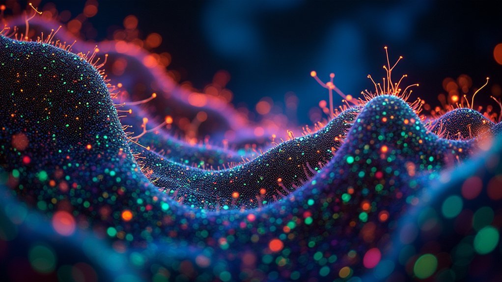
When scientists need to reveal the hidden world within cells, fluorescent stains transform the invisible into spectacular light shows. These remarkable dyes like FITC and rhodamine bind to specific cellular structures, emitting bright colors under UV light for vivid imaging of proteins and organelles.
You’ll find fluorescent stains enable multicolor labeling, where different dyes visualize multiple targets simultaneously, creating richly detailed cellular landscapes.
Using techniques like confocal microscopy, researchers capture high-resolution images that can be reconstructed into three-dimensional structures.
Through the lens of confocal microscopy, the cellular world transforms from flat images into vivid three-dimensional realities.
What’s particularly valuable is live-cell imaging, allowing you to observe cellular functions in real-time—watching division, migration, and transport as they occur.
This technology has revolutionized cancer research and neurobiology, helping identify specific cell types and track responses to treatments, advancing our understanding of complex biological processes.
Gram Staining: The Classic Blue and Pink Contrast
Among the most fundamental techniques in microbiology, Gram staining remains the gold standard for bacterial identification over a century after its development. This differential staining technique creates striking contrasts between gram-positive and gram-negative cells based on their cell wall composition.
When you perform a Gram stain, you’ll witness a beautiful transformation through four key steps:
- Apply crystal violet dye as the primary stain to all bacterial cells.
- Add Gram’s iodine as a mordant to fix the stain.
- Wash with a decolorizing agent (ethanol or acetone).
- Counterstain with safranin, creating the distinctive pink color in gram-negative bacteria.
The resulting blue-purple gram-positive cells and pink gram-negative bacterial pathogens not only create visually compelling images but provide critical information for rapid clinical diagnosis.
Differential Interference Contrast: Revealing Natural Beauty
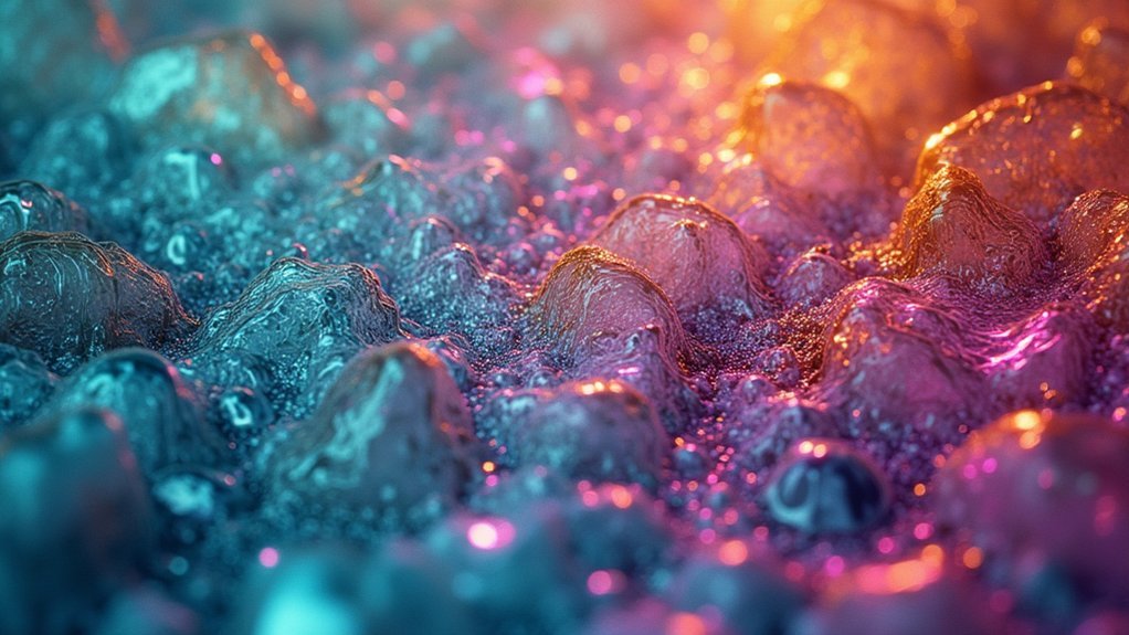
While Gram staining revolutionized our ability to identify bacteria through vibrant color contrasts, Differential Interference Contrast (DIC) microscopy takes a different approach by revealing the natural beauty of living specimens without any staining at all.
When you observe DIC microscopy images, you’ll notice their striking three-dimensional appearance. This technique generates high-contrast images by exploiting differences in cellular structures’ refractive indices, highlighting organelles, membranes, and cytoskeletal elements in their natural state.
You’re fundamentally seeing cells as they truly exist.
Witnessing cellular reality unfiltered, without artificial enhancement or interference – pure biological truth revealed through light.
For researchers studying live cells and their dynamic processes, DIC microscopy offers significant advantages over traditional staining techniques. It preserves cellular integrity while revealing intricate morphology and function.
This makes it invaluable in biological research, especially when observing delicate specimens where stains might introduce unwanted artifacts or alter natural cellular behaviors.
Romanowsky Stains for Blood Cell Visualization
Romanowsky stains transform ordinary blood smears into detailed maps where you’ll immediately recognize different leukocytes by their distinctive coloration patterns.
You can quickly distinguish lymphocytes, neutrophils, and monocytes based on their unique nuclear shapes and cytoplasmic characteristics, making these stains invaluable in hematology labs.
These reliable staining methods enable rapid clinical diagnoses of conditions ranging from infections to leukemias, often providing critical information within minutes of sample preparation.
Differential Blood Cell Identification
When examining blood samples under a microscope, you’ll find that Romanowsky stains provide exceptional visual differentiation of cellular components.
These stains exploit differential staining properties to reveal distinct cell morphology in both white blood cells and red blood cells.
For ideal results with Romanowsky stains, you’ll need to:
- Prepare thin, even blood smears that allow proper fixation
- Identify neutrophils by their multi-lobed nuclei and fine cytoplasmic granules
- Distinguish eosinophils by their bright orange-red granules versus basophils with deep purple-blue granules
- Recognize lymphocytes (round nuclei with minimal cytoplasm) and monocytes (kidney-shaped nuclei with abundant cytoplasm)
The remarkable color variations produced by these stains transform ordinary blood smears into detailed diagnostic landscapes, enabling you to detect abnormalities in cellular components that might indicate hematological disorders.
Fast Clinical Diagnosis Applications
The speed of clinical diagnosis often determines treatment outcomes, particularly in emergency settings. Romanowsky stains serve as powerful tools in rapid diagnosis by revealing critical cellular structures within minutes. When you examine blood smears under the microscope, these stains highlight different cell types through distinctive staining characteristics.
| Condition | Cell Abnormalities | Diagnostic Value |
|---|---|---|
| Leukemia | Immature white cells | Immediate cancer detection |
| Malaria | Parasites in RBCs | Rapid infection confirmation |
| Sepsis | Toxic granulation in neutrophils | Quick infection assessment |
| Thrombocytopenia | Reduced platelets | Urgent bleeding risk evaluation |
The ability to differentiate granulocytes and identify abnormal morphology allows clinicians to initiate treatment within hours rather than days. This rapid clinical diagnostics approach dramatically improves patient outcomes, especially in infectious and hematological emergencies.
Acid-Fast Staining: Highlighting Mycobacteria in Vivid Red
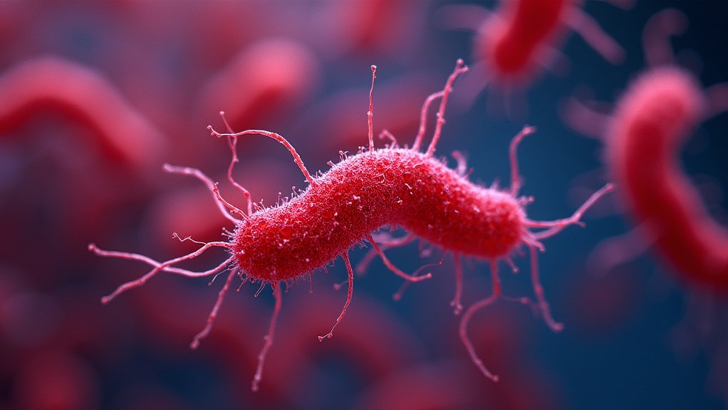
Identifying mycobacteria in clinical samples requires specialized techniques due to their unique cell wall composition. Acid-fast staining uses carbolfuchsin to penetrate and bind to mycolic acids, creating a striking contrast that transforms tuberculosis diagnosis through powerful microscopy.
When you examine these preparations, you’ll notice:
- The vivid red coloration of acid-fast bacteria against a blue background
- Enhanced visualization through the Ziehl-Neelsen method’s heat application
- Clear distinction between mycobacterial presence and other microorganisms
- Detailed morphological features revealed through the stark contrast of stains
The decolorization process washes away carbolfuchsin from non-acid-fast cells, which then absorb the methylene blue counterstain.
This dramatic color difference not only enables rapid identification of potential pathogens but also creates some of the most visually impactful images in clinical microbiology.
Negative Staining Techniques for Capsule Photography
Unlike the vivid colors of acid-fast staining, negative staining techniques reveal what isn’t there rather than what is. When you’re trying to visualize bacterial capsules, traditional positive stains fall short because polysaccharide capsules don’t readily absorb dyes. Instead, India ink or nigrosin creates a dark background while leaving capsules colorless, producing striking halos around cells.
This approach dramatically improves visibility of encapsulated pathogens like Cryptococcus neoformans, where capsule identification can be critical for diagnosis. You’ll notice the capsules appear as transparent rings against the darkened field, creating a photographic negative effect that highlights their presence.
An added benefit of negative staining is its accurate representation of cellular morphology, as it avoids the distortion commonly associated with fixation methods. The result is a clear, precise image that showcases the true structure of these microbial features.
H&E Staining: The Gold Standard for Tissue Imaging
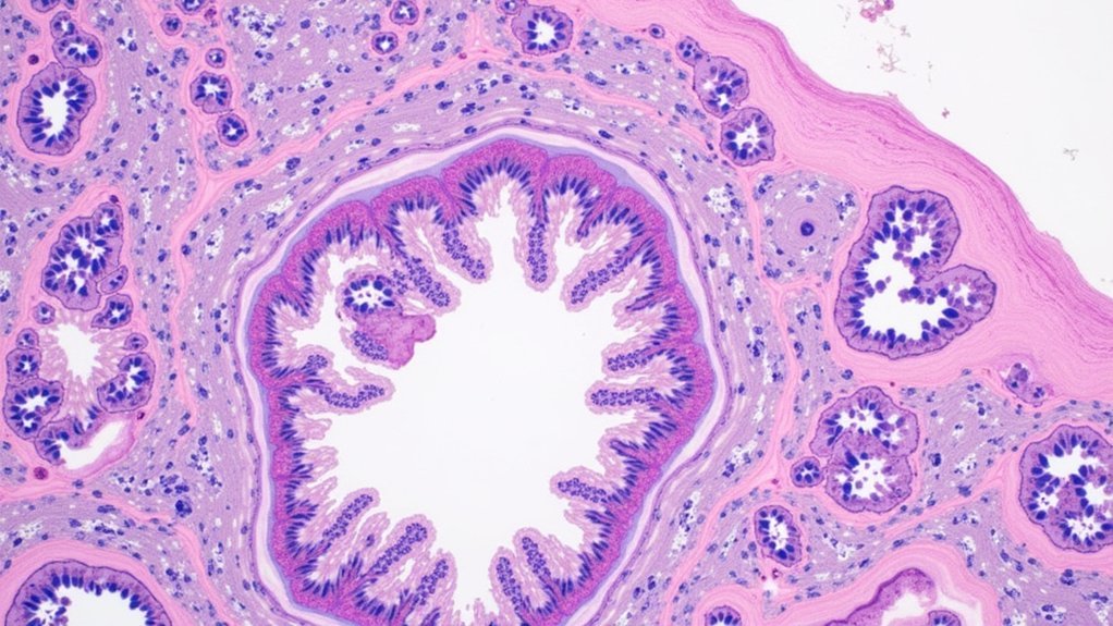
When examining tissue samples in modern histology, you’ll find hematoxylin and eosin (H&E) staining stands firmly as pathology’s most indispensable technique.
This powerful method transforms invisible cellular structures into vibrant, distinguishable elements that reveal critical diagnostic information.
- Dual-color clarity – Hematoxylin binds to nucleic acids, turning nuclei blue, while eosin highlights cytoplasm in contrasting pink or red tones.
- Enhanced tissue definition – The color combination provides exceptional contrast that makes identifying pathologies considerably easier.
- Universal application – You’ll get reliable results across virtually all tissue samples, making it histology’s true gold standard.
- Cost-effective precision – Despite its simplicity, H&E staining delivers remarkable diagnostic accuracy without requiring expensive equipment.
Endospore Stains: Capturing Bacterial Resilience
While H&E staining excels in tissue visualization, the microbiology lab requires specialized techniques to reveal bacterial survival structures. Endospore staining, particularly the Schaeffer-Fulton method, creates striking microscopic images that showcase bacterial resilience at its finest.
Specialized staining reveals the hidden architectural marvels of bacterial endospores, where resilience meets microscopic beauty.
When you examine these preparations, you’ll see vibrant green endospores contrasted against pink vegetative cells, revealing the remarkable dormant structures of Bacillus and Clostridium genera. The process relies on heat fixation to force malachite green through the tough protective layers of endospores, followed by safranin counterstaining.
This visualization technique isn’t just aesthetically pleasing—it’s essential for identifying dangerous pathogens like B. anthracis and C. difficile.
Microbiologists routinely use these staining techniques to rapidly identify spore-forming bacteria, offering immediate insights into their survival mechanisms and pathogenic potential.
DAPI and Nuclear Visualization Techniques
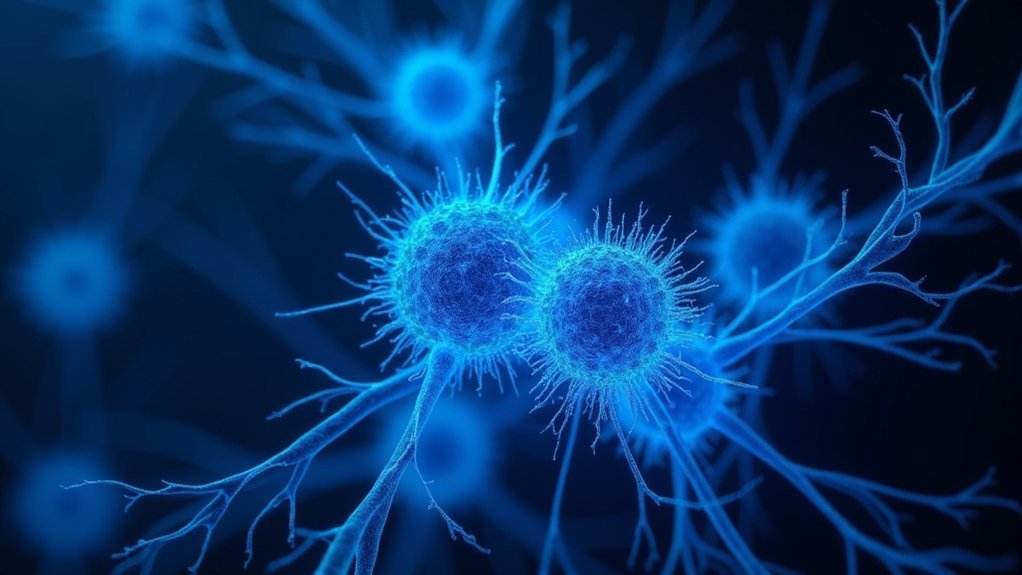
Beneath the brilliant blue glow of ultraviolet light, DAPI transforms ordinary cellular samples into cosmic-like landscapes where nuclei shine like stars against the darkness. This powerful fluorescent stain binds specifically to A-T rich regions in DNA, revealing nuclear morphology with exceptional clarity.
When you’re seeking stunning biological specimens, DAPI offers unique advantages:
- Speed and simplicity – Complete staining in just 5-10 minutes without compromising cellular integrity
- Exceptional contrast – Visualize nuclei with vivid blue fluorescence against dark backgrounds
- Cell cycle visualization – Identify different stages of mitosis and apoptosis with remarkable detail
- Multiplexing capability – Combine with other fluorescent markers for multi-channel imaging techniques that showcase cellular structures simultaneously
DAPI’s integration with fluorescence microscopy creates images that are both scientifically valuable and visually enchanting.
Immunofluorescence: Targeting Specific Structures With Precision
Immunofluorescence takes cellular visualization beyond simple DNA binding to illuminate the intricate molecular landscape within biological specimens. You’ll discover how this technique uses fluorescent dyes attached to antibodies that precisely target specific proteins, revealing their spatial distribution with remarkable clarity.
What makes immunofluorescence particularly powerful is your ability to visualize multiple cellular processes simultaneously by using different colored fluorescent markers. This multiplexing approach discloses complex biological structures and their interactions at the molecular level.
From identifying pathogens to studying cancer markers, immunofluorescence applications span both research and clinical diagnostics.
When you need even greater detail, super-resolution techniques can enhance these images to uncover nanoscale structures previously hidden from view. The result? Stunningly detailed maps of cellular architecture that transform our understanding of biological function.
Silver Staining Methods for Nerve Tissue and Fungi
Silver staining techniques represent a crucial advancement in the visualization of neural architecture and fungal structures that are otherwise difficult to observe with conventional stains.
You’ll find these methods particularly valuable in histopathology for diagnosing conditions involving delicate tissue components.
- Nerve tissue visualization – Gomori’s silver stain precipitates silver chromate around proteins, creating brown or black outlines that reveal the intricate morphology of axons and dendrites.
- Fungal studies – Grocott’s methenamine silver targets polysaccharide components in fungal cell walls, enabling clear differentiation from surrounding tissues.
- Connective tissue examination – Silver stains highlight reticular fibers, exposing the complex supporting framework of tissue architecture.
- Enhanced sensitivity – These methods detect low-abundance structures that conventional stains might miss, making them indispensable for neural and fungal pathology.
PAS Staining for Colorful Fungal and Tissue Documentation
While silver staining techniques excel at revealing neural structures and fungi, the Periodic Acid-Schiff (PAS) method offers a distinct advantage through its vibrant color profile.
You’ll immediately notice the striking magenta or pink hues that highlight polysaccharides when viewing PAS-stained specimens under your microscope.
This technique creates exceptional contrast that makes fungal cell walls pop against surrounding tissues. When you’re documenting pathological specimens, PAS staining reveals glycogen accumulation in liver samples or abnormal mucin production in certain cancers with remarkable clarity.
The process involves oxidizing tissue polysaccharides with periodic acid before applying Schiff’s reagent, creating that characteristic colorful reaction.
For both diagnostic and research purposes, you’ll find PAS staining invaluable when you need to visualize and document carbohydrate-rich structures in biological samples.
Digital Coloration Techniques for Enhanced Microscopic Imagery
Digital coloration has evolved beyond traditional staining with AI color remapping now transforming grayscale microscopic images into vibrant, interpretable visualizations.
You’ll find these computational techniques can highlight structures that would otherwise remain invisible, assigning specific colors to different spectral signatures within your samples.
Spectral analysis enhancement further refines this approach by separating overlapping fluorescent signals, allowing you to distinguish multiple cellular components simultaneously with unprecedented clarity.
AI Color Remapping
As microscopy continues to evolve, AI color remapping has emerged as a revolutionary approach to biological imaging. You’ll find that these advanced digital techniques transform traditional staining methods by algorithmically enhancing the colors of biological samples, dramatically improving contrast between cellular structures.
With AI color remapping, you can:
- Highlight specific cellular interactions that would remain hidden with conventional staining
- Adjust color palettes to improve interpretability of complex biological data
- Distinguish between similar cellular components with unprecedented clarity and detail
- Create visually stunning images that serve both scientific and aesthetic purposes
These algorithms analyze microscopic imagery and digitally alter color schemes, allowing you to visualize patterns and anomalies that inform diagnostics and research.
The technology enables you to see beyond the limitations of traditional stains, revealing the hidden complexity of biological specimens.
Spectral Analysis Enhancement
Behind every remarkable biological discovery lies the power of spectral analysis enhancement techniques. When you’re examining microscopic images, digital coloration techniques transform ordinary specimens into revelatory visual data.
By manipulating color channels through advanced imaging software, you’ll highlight specific cellular structures that might otherwise remain hidden. These color-enhanced visuals aren’t just aesthetically pleasing—they’re scientifically essential.
Pseudo-coloring converts grayscale images into vibrant representations where different biochemical properties become instantly distinguishable. You’ll notice subtle variations in cell morphology that standard staining methods simply can’t reveal.
The real magic happens when spectral analysis combines with digital processing. This partnership illuminates the intricate details of microscopic images, allowing you to differentiate between neighboring structures with similar appearances but different compositions.
Your research benefits from both enhanced visibility and more accurate diagnostics.
Frequently Asked Questions
What Are the 3 Types of Biological Stains?
You’ll find three main types of biological stains: positive stains, which color the cells; negative stains, which color the background; and differential stains, which use multiple dyes to distinguish between cellular structures.
Which Characteristic of an Image Does Staining Improve Most?
Staining improves contrast in your biological images the most. You’ll notice cellular structures become considerably more visible against backgrounds, which is essential when you’re analyzing tissues or microorganisms under the microscope.
Which Type of Stain Best Stains the Bacterial Cell?
The Gram stain is your best option for bacterial cells as it’ll clearly differentiate between Gram-positive and Gram-negative bacteria based on their cell wall composition, revealing their fundamental structural characteristics.
Why Are Most Biological Stains Positively Charged?
Biological stains are positively charged because they’ll bind effectively to your cell’s negatively charged components like nucleic acids and proteins. This attraction creates better visibility and contrast when you’re examining cellular structures under the microscope.
In Summary
You’ll find that the world of biological staining is as artistic as it is scientific. Whether you’re working with fluorescent dyes that transform cells into neon landscapes or classical Gram stains that reveal bacterial identities, each technique offers a unique window into the microscopic domain. Choose your staining method based on your specimen and what you’re hoping to reveal—the most stunning images often emerge when technique meets curiosity.



Leave a Reply