For time-lapse research, you’ll find exceptional performance from Incucyte Live-Cell Analysis Systems offering real-time monitoring without disruption, ImageXpress High-Content Platforms with dynamic visualization capabilities, Agilent BioTek Cytation Series combining microscopy with microplate detection, CrestOptics DeepSIM providing super-resolution at 100nm, and Light Sheet Microscopy Systems that reduce phototoxicity for extended observations. Each system delivers unique advantages for capturing cellular processes as they unfold over time.
Incucyte Live-Cell Analysis Systems for Continuous Monitoring
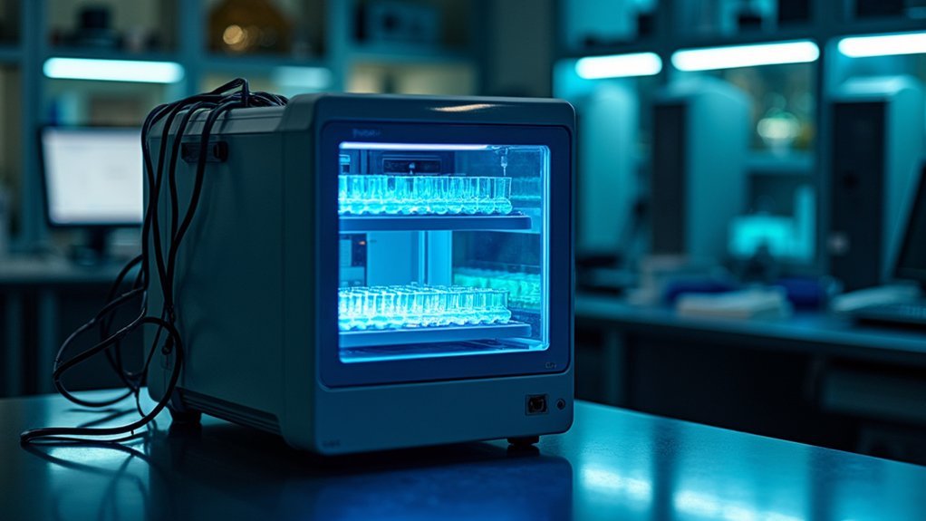
When researchers need to track cellular activities over extended periods, Incucyte Live-Cell Analysis Systems stand out as powerful tools for continuous monitoring.
You’ll benefit from real-time monitoring of cellular behavior without disrupting your experiments, allowing you to observe responses as they occur naturally.
The flagship Incucyte S3 excels in viral studies and apoptosis analysis, making it ideal for larger laboratories.
If you’re focused on cell proliferation and transfection efficiency, the SX1 offers automated image acquisition and analysis features that boost your research productivity.
For metabolic studies requiring exceptional imaging quality, the SX5’s patent-pending optics deliver superior results.
All systems provide high-content imaging capabilities across diverse biological research applications, ensuring you’ll capture extensive data for thorough analysis of dynamic cellular processes.
ImageXpress High-Content Platforms for Time-Lapse Research
While tracking cellular dynamics requires sophisticated instrumentation, ImageXpress High-Content Imaging Systems deliver exceptional capabilities for your time-lapse research needs.
The ImageXpress Confocal HT.ai system features a seven-channel laser light source that produces outstanding image quality for cancer research and COVID-19 studies.
For your basic to advanced screening requirements, the ImageXpress Micro 4 offers versatile widefield fluorescence microscopy with robust time-lapse capabilities.
If you’re conducting RNA interference or stem cell studies, you’ll find the ImageXpress Nano particularly user-friendly, providing accessible high-content imaging.
Each platform excels in live-cell imaging, allowing you to monitor cellular processes in real-time.
These systems combine exceptional cell analysis tools with time-lapse functionality, making them indispensable for researchers who need to visualize dynamic biological events.
Agilent BioTek Cytation Series: Versatility in Time-Lapse Applications
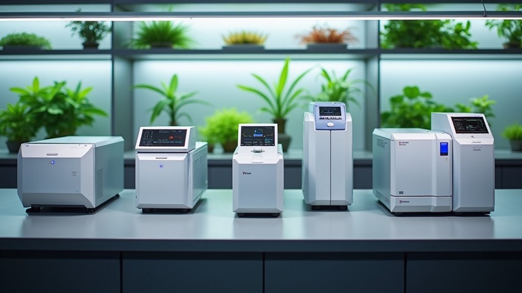
The Agilent BioTek Cytation Series stands as another powerful option in your time-lapse imaging toolbox, offering a distinctive combination of automated microscopy with conventional microplate detection.
Sophisticated yet intuitive, the Cytation Series merges automated microscopy with microplate detection for unparalleled time-lapse imaging capabilities.
You’ll appreciate how these robust imaging systems excel in biological research applications where monitoring cellular processes over time is critical.
- Supports real-time live cell imaging, allowing you to track dynamic changes continuously
- Integrates cell counting and viability assessment features for quantitative time-lapse studies
- Excels in high content screening with efficient data management capabilities
- Delivers high-quality images consistently, essential for accurate time-lapse analysis
- Provides versatility across diverse time-lapse applications, from basic research to complex cellular assays
This integration of technologies makes the Cytation Series particularly valuable when you’re conducting extended experiments requiring both visual and quantitative data collection.
CrestOptics DeepSIM: Super-Resolution Time-Lapse Capabilities
Pushing the boundaries of conventional imaging, CrestOptics DeepSIM delivers extraordinary super-resolution capabilities that transform time-lapse research.
With its multi-spot lattice structured illumination technology, you’ll achieve remarkable 100 nm XY resolution, revealing unprecedented imaging detail in your studies.
When you’re tracking dynamic cellular processes, DeepSIM excels at live-cell imaging, maintaining high fidelity throughout your observations.
The system penetrates thick samples with super-resolved data up to 50 µm deep, allowing you to explore complex biological structures thoroughly.
You’ll appreciate the significant improvements in accuracy for time-lapse microscopy, essential when studying cellular behavior over extended periods.
For researchers demanding high-quality images with exceptional reliability, DeepSIM stands out as an invaluable tool in super-resolution microscopy.
Light Sheet Microscopy Systems for Extended Time-Lapse Imaging
When studying live specimens over extended periods, Light Sheet Microscopy systems revolutionize time-lapse imaging with their unique illumination approach.
These systems offer vital advantages for capturing cellular processes without damaging your valuable samples.
Light sheet technology preserves specimen integrity while delivering unparalleled visibility into dynamic cellular events.
- UltraMicroscope Blaze delivers automated 3D imaging perfect for long-duration time-lapse studies
- Notably reduced phototoxicity and photobleaching lets you observe live specimens for extended periods
- UltraMicroscope II excels in fluorescence imaging of large samples with exceptional detail
- Multi-channel high-resolution imaging capabilities reveal complex biological interactions in real-time
- Fast imaging speeds capture dynamic events that would be missed with conventional microscopy
You’ll find these systems particularly valuable when your research demands both gentle treatment of samples and thorough visualization of cellular processes over time.
Frequently Asked Questions
What Is the Time-Lapse Imaging Technique?
Time-lapse imaging is a technique where you’ll capture images at set intervals, creating a video that reveals slow changes over time. You’re fundamentally compressing hours or days into minutes for visualization.
What Is Time-Lapse Confocal Imaging?
Time-lapse confocal imaging captures sequential images using laser scanning to eliminate out-of-focus light. You’ll see cellular processes unfold with high spatial resolution, allowing you to track dynamic biological changes over extended periods.
What Microscope Can Be Used to View Live Cells?
You can view live cells using several microscopes including Agilent’s BioSpa system, Leica’s STELLARIS STED confocal, Miltenyi’s UltraMicroscope II, and Nikon’s ECLIPSE Ji, all designed for non-disruptive observation of living cells.
What Is Time-Lapse Imaging in IVF?
Time-lapse imaging in IVF lets you monitor embryo development continuously without disturbing them. It captures images at set intervals, showing growth patterns and helping you select the most viable embryos for transfer.
In Summary
Whether you’re monitoring cell cultures over days or capturing rapid subcellular events, you’ll find the perfect fit among these top imaging systems. You’re investing in technology that balances resolution, throughput, and gentle imaging conditions. Whichever platform you choose, you’re now equipped to make an informed decision that’ll advance your time-lapse research with reliable, high-quality data and streamlined workflows.

Leave a Reply