For stunning darkfield microscopy, you’ll need three key instruments: High-contrast darkfield condensers that direct light at oblique angles to illuminate transparent specimens brilliantly; Scientific CMOS cameras with superior sensitivity and resolution to capture fine details in low-light conditions; and Specialized LED light sources that provide precise illumination control for ideal background darkness. These essential tools transform invisible specimens into vibrant subjects while revealing structures that traditional methods miss. Discover how these instruments can revolutionize your microscopic imaging capabilities.
High-Contrast Darkfield Condensers: Maximizing Specimen Illumination
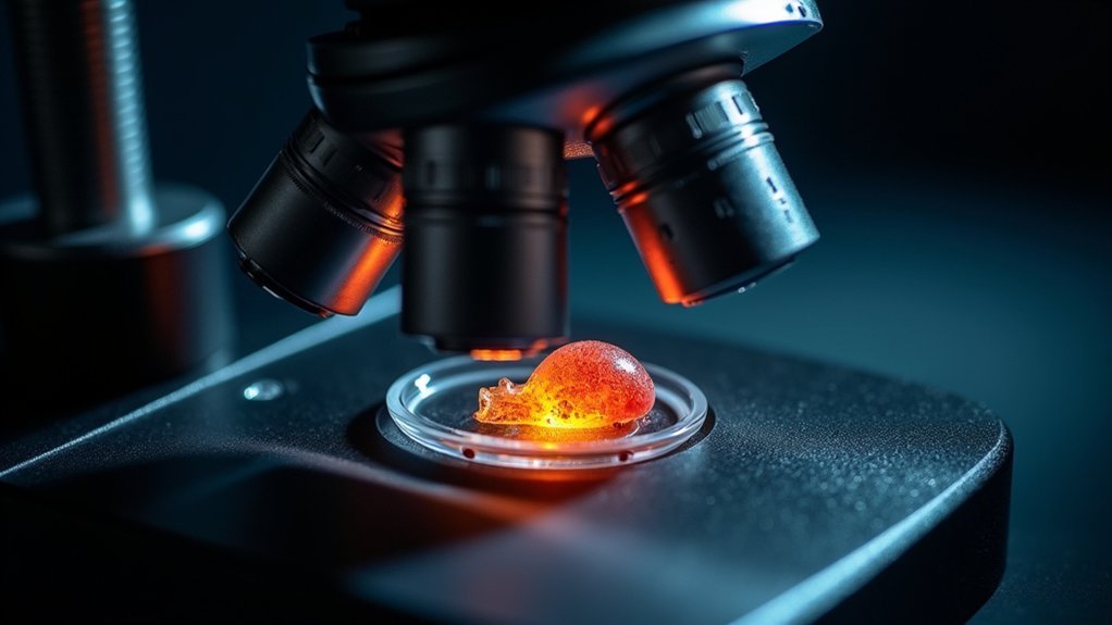
Three critical features make high-contrast darkfield condensers indispensable tools for capturing stunning microscopic images.
First, they direct light at oblique angles, creating maximized illumination of transparent organisms that would otherwise appear invisible.
Second, their specialized opaque disk blocks direct light while allowing only scattered light from specimens to reach your objective lens, dramatically enhancing contrast.
You’ll achieve superior results by adjusting condenser height and aperture diaphragm settings to fine-tune illumination levels for your specific specimen.
This precision control reveals delicate structures in bacteria and protozoa that standard brightfield microscopy simply can’t detect.
In clinical diagnostics, these condensers prove particularly valuable, enabling identification of pathogens like Treponema pallidum without traditional staining.
Darkfield microscopy with high-quality condensers transforms invisible specimens into brilliantly illuminated subjects against a black background.
Scientific CMOS Cameras: Capturing Detail in Low-Light Conditions
Scientific CMOS cameras represent a revolutionary advancement in darkfield microscopy, delivering exceptional performance when light levels would typically limit image quality.
You’ll achieve superior results with these cameras’ high sensitivity capabilities, allowing you to maximize illumination even in the most challenging viewing conditions.
With resolutions exceeding 1 megapixel, scientific CMOS cameras enable detail visualization of structures that conventional cameras simply miss.
Their advanced noise reduction technology enhances contrast while maintaining clarity—essential for darkfield microscopy’s unique lighting challenges.
Perhaps most impressively, these cameras capture images at high frame rates, making them ideal for observing dynamic live specimens in real time.
You’ll appreciate the fast readout speeds that don’t compromise image quality, ensuring you never miss critical moments in your darkfield microscopy work.
Specialized LED Light Sources: Achieving Optimal Background Darkness
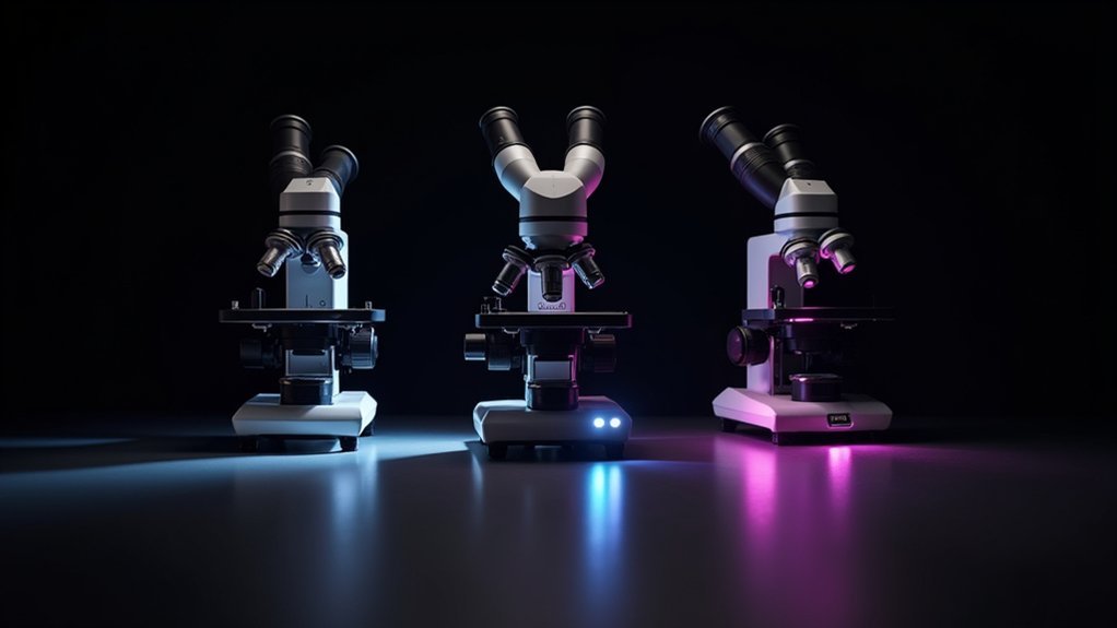
Modern darkfield microscopy demands specialized LED light sources that deliver precise illumination control. You’ll achieve stunning contrast between unstained specimens and a dark background with these high-intensity light systems. Their adjustable intensity settings let you fine-tune illumination to reveal delicate structures while minimizing scatter.
| Feature | Benefit |
|---|---|
| Specific wavelengths | Reduces scatter for clearer visualization |
| Adjustable intensity | Eliminates glare for better image quality |
| Uniform illumination | Creates consistent dark backgrounds |
| Low heat output | Enables longer imaging sessions |
| System compatibility | Offers versatility with existing setups |
Unlike traditional halogen bulbs, these specialized LED light sources run cooler and last considerably longer, making them ideal for extended observation sessions. Their ability to highlight minute details in biological specimens transforms ordinary observations into enchanting scientific imagery.
Frequently Asked Questions
What Is the Dark Field Microscope Instrument?
A dark field microscope is your specialized optical instrument that blocks direct light to create bright images of specimens against a dark background, allowing you’ll see transparent organisms without staining.
What Instrument Produces a Bright Image of the Specimen Against a Dark Background?
Darkfield microscopes produce the bright image of specimens against a dark background. You’ll need one with a specialized darkfield condenser that blocks direct light and only allows scattered light to reach your objective lens.
What Instrument Is Used to See a Highly Magnified Image of an Object?
You’ll need a compound microscope to see highly magnified objects. It uses multiple lenses – an objective and eyepiece – to achieve magnifications from 40× to 1000×, revealing details invisible to the naked eye.
Which of the Following Instruments Is Best Used to Create a Three Dimensional Image of a Cell’s Organelle?
For creating three-dimensional images of cell organelles, you’ll want to use a confocal microscope. It scans multiple z-planes with lasers, allowing you to visualize the complete spatial arrangement of organelles within cells with high resolution.
In Summary
You’ve now discovered the three essential tools for capturing breathtaking darkfield images. By investing in a high-contrast darkfield condenser, you’ll dramatically enhance specimen illumination. Pair this with a scientific CMOS camera that excels in low-light environments, and add specialized LED light sources for perfect background darkness. With this powerful combination, you’ll create striking images that reveal microscopic details invisible to conventional microscopy techniques.


Leave a Reply