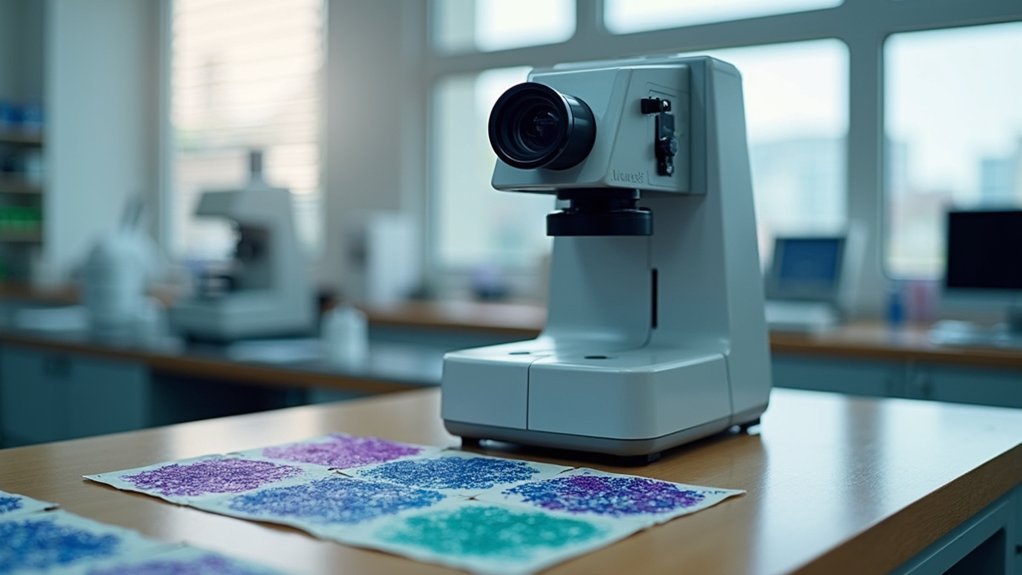
When selecting digital pathology cameras, prioritize high resolution (0.172 µM/pixel), scanning speed (60-100+ fps), and seamless integration with your existing laboratory systems. Look for models supporting standard formats like DICOM and SVS, with automatic quality control features. Budget considerations should include long-term costs (maintenance runs 15-20% of purchase price annually). Top performers include Huron’s TissueScope™ iQ and Leica’s Aperio GT 450 DX. Continue exploring to discover which camera specifications will transform your diagnostic workflow.
8 Second-Level Headings for “Top Digital Pathology Cameras: Complete Buyer’s Guide”

To craft an effective buyer’s guide for digital pathology cameras, you’ll need seven essential section headings that guide readers through the selection process.
Finding the right digital pathology camera requires strategic evaluation of key features that align with clinical demands and workflow requirements.
First, include “Understanding Digital Pathology Technology” to establish foundational knowledge.
Follow with “Image Quality Essentials” covering resolution and color accuracy critical for diagnosis.
Add “Scanning Speed and Efficiency” to address how faster cameras improve workflow efficiency.
Include “Compatibility and Integration” discussing how cameras work with existing systems.
“File Format Support” should cover SVS, DICOM, and other formats.
Add “User-friendly Interface Features” focusing on control software and accessibility.
Finally, include “Price-to-Performance Analysis” to help readers balance budget constraints with technical requirements.
These headings create a thorough framework for evaluating digital pathology cameras that enhance accuracy and efficiency in clinical settings.
Essential Features of High-Performance Digital Pathology Cameras
Precision and reliability form the backbone of any high-performance digital pathology camera system. When selecting your equipment, focus on spectral sensitivity range—top models cover 400-750nm with extensions into UV and NIR for specialized applications.
You’ll want cameras designed for low-light conditions that can detect as few as 1-100 photons per pixel. Look for advanced features like global shutter technology that minimizes motion artifacts by capturing the entire pixel array simultaneously, dramatically enhancing image quality.
High frame rates exceeding 100fps make dynamic imaging applications and real-time analysis possible.
Don’t overlook cooling systems that reduce dark current noise—these enable long exposure imaging lasting from seconds to hours without compromising clarity.
These technical capabilities directly impact diagnostic accuracy and workflow efficiency in your digital pathology practice.
Resolution and Image Quality Considerations for Pathology Applications

When diagnosing complex tissue samples, resolution becomes the critical factor that determines diagnostic accuracy and confidence. Your digital pathology camera should deliver high-resolution imaging at 0.172 µM/pixel to reveal intricate cellular structures invisible to lower-quality systems.
| Resolution Factor | Impact on Diagnosis |
|---|---|
| Magnification (1.25x-100x) | Enables visualization from macro to subcellular level |
| Scanning Method | Automatic narrow stripe scanning preserves critical details |
| Slide Size Support | Accommodates specimens from 15mm² to 208mm x 75mm |
| Image Format | SVS and NDPI formats guarantee universal compatibility |
| Quality Control Algorithms | Verify imaging accuracy for reliable clinical results |
The best pathology applications rely on cameras that balance resolution capabilities with proven quality control systems, making sure you’ll never miss subtle pathological changes that could affect patient outcomes.
Speed and Throughput: Balancing Efficiency With Precision
While high resolution remains foundational to diagnostic accuracy, the speed at which a digital pathology camera operates defines laboratory throughput and overall efficiency.
Today’s high-speed CMOS models exceed 100 fps, dramatically improving workflow in busy labs.
You’ll find significant variation in scanning speeds across systems. Huron’s TissueScope™ iQ averages 60 seconds per slide, while Grundium’s Ocus®20 prioritizes rapid processing for time-sensitive applications.
For high-volume operations, Leica’s Aperio GT 450 DX offers no-touch scanning for 450 slides, balancing throughput with user-friendly operation.
The integration of automated quality control features, like those in MoticEasyScan’s technology, helps maintain precision while accelerating slide management.
When selecting digital pathology equipment, consider how these speed capabilities align with your laboratory’s specific throughput requirements.
Compatibility and Integration With Existing Laboratory Systems

Successful implementation of digital pathology cameras hinges on their ability to integrate seamlessly with your laboratory’s existing infrastructure. When evaluating options, prioritize systems that support standard file formats like DICOM and SVS to guarantee compatibility with your laboratory information management systems.
- Look for cameras that connect effortlessly to your existing IT infrastructure for streamlined data transfer.
- Verify support for third-party image analysis software like QuPath and Indica Labs.
- Check the vendor’s integration history with established LIMS providers.
- Guarantee robust cybersecurity features for protecting patient data.
- Confirm that workflow efficiency won’t be compromised during implementation.
Remember that even the highest resolution camera won’t deliver value if it creates integration bottlenecks or requires complete system overhauls. The right digital pathology camera should enhance your current operations, not disrupt them.
Budget Planning: Upfront Costs vs. Long-Term Value
Budget planning for digital pathology cameras extends beyond mere compatibility considerations into hard financial decisions.
You’ll face initial budget allocation challenges with upfront costs ranging from $10,000 for basic models to over $40,000 for advanced systems.
When evaluating long-term value, factor in annual maintenance expenses (15-20% of purchase price), training costs, and integration costs that guarantee your team maximizes the technology’s potential.
Consider how higher-quality cameras can enhance diagnostic accuracy, potentially improving patient outcomes while reducing expenses associated with misdiagnoses.
To make informed decisions, calculate the total cost of ownership over 5-10 years.
This all-encompassing approach helps balance immediate expenditure against future savings, guaranteeing your digital pathology systems deliver peak value throughout their lifecycle rather than just meeting immediate budgetary constraints.
Key Technical Specifications That Impact Diagnostic Accuracy

Image quality serves as the foundation of accurate pathological diagnosis in the digital domain.
Image quality forms the critical bedrock upon which precise digital pathological interpretations depend.
When selecting digital pathology cameras, you’ll need to evaluate several technical specifications that directly influence diagnostic accuracy:
- Spectral range (400-750 nm) – Broader ranges with UV/NIR capabilities reveal subtle tissue features invisible to standard cameras.
- Low dark current – Essential for extended exposure imaging, reducing noise in long-duration captures of static samples.
- Frame rate capabilities – High-speed CMOS models (>100 fps) capture dynamic samples with exceptional temporal resolution.
- Sensitivity metrics – Cameras with SNR above 1.80 at five photons per pixel detect minute cellular details.
- Quantum efficiency – Higher efficiency translates to better low-light performance, critical for identifying faint structures in tissue samples.
Software Solutions for Image Management and Analysis
The power of digital pathology cameras can only be fully realized when paired with robust software solutions that efficiently manage and analyze the wealth of data they generate.
When selecting your digital pathology system, you’ll need image management systems that seamlessly integrate with your existing LIS while supporting diverse file formats like SVS, NDPI, and TIFF.
Look for analysis tools that facilitate annotation and manipulation of high-resolution digital images, including features for regions of interest, Z-stack, and TMA analysis to enhance diagnostic accuracy.
Automation features such as barcoding streamline workflow processes and guarantee sample tracking.
For managing large data volumes, prioritize solutions with cloud storage compatibility.
Advanced software incorporating AI algorithms can dramatically improve efficiency by automating routine tasks and enhancing specific cell identification within your digital images.
Frequently Asked Questions
How Long Is the Typical Lifespan of a Digital Pathology Camera?
Digital pathology cameras typically last 5-7 years with proper maintenance. You’ll get the longest lifespan if you’re handling them carefully, keeping them clean, and following manufacturer’s guidelines for usage and storage.
Can These Cameras Be Used for Research Beyond Clinical Diagnostics?
Yes, you’ll find these cameras extremely valuable for research beyond diagnostics. They’re perfect for specimen documentation, educational purposes, biomarker studies, and collaborative research where high-resolution imaging and detailed analysis are essential.
What Certifications Should I Look for When Purchasing?
Look for cameras with FDA approval for clinical use, CE marking for European markets, ISO 13485 certification, and CLIA compliance. Don’t forget connectivity certifications like DICOM if you’ll integrate with hospital systems.
How Do Storage Requirements Scale With Increased Camera Usage?
Storage requirements grow exponentially with increased camera usage. You’ll need 1-3TB per month for standard use, but this can triple with higher resolution cameras or additional imaging sessions. Consider cloud solutions for scalability.
Are There Significant Differences Between Cameras for Teaching Versus Diagnosis?
Yes, there are significant differences. For teaching, you’ll need cameras with good sharing capabilities but lower resolution. For diagnosis, you’ll require higher resolution, color accuracy, and FDA-approved equipment that guarantees diagnostic reliability.
In Summary
When choosing your digital pathology camera, prioritize features that match your specific workflow needs. You’ll find the perfect balance between resolution, speed, and compatibility makes all the difference in diagnostic accuracy. Remember that initial investment should be weighed against long-term value. With the right camera and software combination, you’re not just buying equipment—you’re investing in your lab’s diagnostic future.


Leave a Reply