Looking for top microscope imaging software? Start with Leica Application Suite (LAS X) for extensive control, ZEN (Zeiss) for intuitive workflows, or Imaris for advanced 3D/4D visualization. NIS-Elements offers unified control across different microscopes, while open-source options include ImageJ/Fiji and CellProfiler. Each tool brings unique strengths to your research, from high-throughput analysis to specialized visualization. Discover which platform best matches your specific imaging needs below.
7 Top Microscope Imaging Software Tools to Try

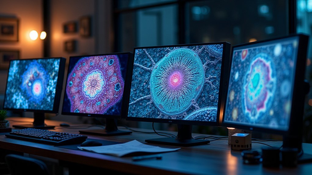
Today’s microscope imaging software tools frequently offer much more than basic image capture capabilities.
If you’re looking for extensive image analysis software, consider Leica Application Suite (LAS X), which integrates microscope and camera control with specialized modules for diverse scientific applications.
For advanced 3D/4D visualization, Imaris provides powerful analysis tools with packages tailored to specific research requirements.
NIS-Elements offers a unified interface across multiple imaging systems with customizable hardware selection and remote monitoring features.
Budget-conscious researchers can turn to free options like Fiji and CellProfiler for fluorescent imaging analysis, quantification, and 3D visualization.
Free tools like Fiji and CellProfiler offer comprehensive solutions for researchers needing powerful imaging analysis without the price tag.
Meanwhile, platform-specific solutions include Enersight for Windows users and Leica Acquire for Mac environments, ensuring you’ll find suitable imaging solutions regardless of your operating system preference.
Leica Application Suite X (LAS X): Integrated Microscopy Platform
Leica Application Suite X streamlines your microscopy workflow by integrating microscope control and camera management in one interface.
The software’s modular architecture lets you choose specialized components for Life Science, Industry, or Materials Science while providing customizable report templates and advanced analysis tools.
You’ll need to evaluate platform compatibility when implementing LAS X, though extensive learning resources including tutorials and YouTube videos help maximize your imaging capabilities regardless of your setup.
Integrated Workflow Systems
When researchers need extensive control over their microscopy workflow, the Leica Application Suite X (LAS X) stands out as a powerful integrated platform. This software platform seamlessly combines microscope and camera control, creating a streamlined imaging experience tailored for both Life Science and Industry applications.
You’ll appreciate LAS X’s specialized modules that enable full-screen observation, enhanced image stitching, and customizable report templates. The LAS X Navigator provides quick overviews of your specimens, simplifying complex imaging tasks while integrating various Leica instruments into your workflow.
The system’s automatic calibration adds professional scale bars to your images, while basic annotation tools let you incorporate essential data.
With thumbnail galleries for efficient image review and flexible exposure controls, you’ll achieve reliable results regardless of your application needs.
Modular Application Architecture
The modular architecture of LAS X represents the cornerstone of its versatility and power across diverse microscopy applications. This extensive software package adapts to your specific research needs through specialized modules for Life Science, Industry, and Materials Science—each delivering tailored analysis tools and reporting capabilities.
You’ll appreciate how LAS X streamlines your workflow with its intuitive thumbnail gallery for quick image review and automatic scale bar calibration that guarantees measurement accuracy.
Whether you prefer auto or manual exposure adjustments, the platform delivers consistently reliable imaging results across different microscopy techniques.
What makes LAS X particularly valuable is its seamless compatibility with previous Leica Application Suite versions, allowing you to upgrade without sacrificing functionality while gaining access to advanced imaging features that enhance your research capabilities.
Platform Compatibility Considerations
Operating your LAS X software requires specific system prerequisites that guarantee peak performance during complex imaging tasks. You’ll need either Windows 10 Pro or Windows 11, as LAS X is exclusively designed for Windows environments.
When setting up for image acquisition, remember that licensing requires a hardware dongle—either as a soft dongle installed directly on your PC or a physical USB stick. This guarantees proper authentication while allowing flexibility in how you manage your license.
The software seamlessly integrates with all Leica microscopes and digital cameras, creating a unified workflow environment.
Whether you’re working in Life Science, Industry, or Materials Science, you’ll find dedicated modules optimized for your specific imaging needs.
LAS X also maintains compatibility with previous Leica Application Suite versions, making your shift smooth and preserving access to earlier projects.
Imaris: Advanced 3D/4D Visualization and Analysis
Renowned for transforming the way scientists explore complex biological structures, Imaris stands as a premier software solution for advanced 3D/4D visualization and analysis.
You’ll find its Super Resolution capabilities particularly valuable when examining intricate cellular components that traditional imaging methods can’t capture effectively.
The software offers tailored packages designed for specific research applications, with an intuitive interface that streamlines complex tasks.
Imaris Infinity pushes visualization boundaries, providing sophisticated tools that enhance your analytical capabilities across multiple scientific disciplines.
Before committing, you can test Imaris through trial periods to experience its market-leading functionalities firsthand.
With regular updates introducing cutting-edge features, you’ll maintain access to the latest advancements, ensuring your research remains at the forefront of imaging technology.
NIS-Elements: Unified Interface for Multiple Imaging Systems
While Imaris excels in 3D visualization, NIS-Elements represents Nikon’s extensive approach to microscopy software integration.
You’ll appreciate how it provides consistent control across widefield, confocal, and super resolution image systems through a single interface.
NIS-Elements allows you to effortlessly switch between different microscope platforms and combine results from various Nikon systems, streamlining your research workflow.
The software’s customization features let you tailor hardware selection and acquisition routines to match your specific experimental needs.
Data export is hassle-free with support for multiple formats and software platforms, enhancing collaboration possibilities.
You can even operate and monitor your microscopes remotely without installing software on off-site computers—perfect for troubleshooting experiments from anywhere.
ImageJ/Fiji: Open-Source Analysis Powerhouse
ImageJ/Fiji stands as the cornerstone of open-source image analysis software in scientific research, offering you unprecedented flexibility without licensing costs. This powerful platform excels at handling diverse image formats like TIFF, JPEG, and PNG, while effortlessly managing multi-dimensional data for 3D visualization.
You’ll benefit from advanced features including z-projection, segmentation, and particle analysis—essential tools for quantifying fluorescence and other image characteristics. Fiji enhances the ImageJ experience by arriving pre-packaged with specialized plugins tailored for biological research.
What truly sets this software apart is its vibrant community support, providing continuous updates and extensive online resources.
Whether you’re processing simple micrographs or complex image stacks, ImageJ/Fiji delivers the customizable workflows you need for sophisticated scientific image analysis.
CellProfiler: Automated Image Processing for High-Throughput Data
CellProfiler empowers you to analyze thousands of images automatically through its robust high-throughput capabilities, saving you countless hours of manual work.
You’ll find creating customized processing pipelines straightforward using the intuitive drag-and-drop interface, even without programming expertise.
As a free open-source platform, CellProfiler offers research labs of all sizes access to sophisticated image analysis tools without budget constraints.
High-Throughput Analysis Capabilities
Modern biomedical research often generates thousands of microscopy images that require efficient processing and analysis. CellProfiler excels in this domain, giving you powerful tools to handle massive datasets without programming expertise.
With CellProfiler’s high-throughput capabilities, you’ll advance your research through:
- User-friendly pipeline building interface for customizable analysis workflows
- Support for multiple image formats and multi-channel analysis across diverse biological applications
- Built-in measurements for cell morphology, intensity, and texture providing extensive quantification
- Seamless integration with high-content screening systems
- Automated processing that transforms hours of manual work into efficient, reproducible analysis
You can quickly set up analysis pipelines that process entire experimental datasets, extract meaningful measurements, and generate quantitative results that support data-driven conclusions in your advanced research projects.
Customizable Processing Pipelines
The heart of CellProfiler’s analytical power lies in its customizable processing pipelines, which transform complex image analysis tasks into streamlined workflows. You’ll find creating these pipelines intuitive through the graphical user interface, eliminating the need for extensive programming knowledge.
Each pipeline consists of carefully arranged modules that perform specific functions—from image enhancement and segmentation to quantification. You can easily modify existing pipelines or build new ones tailored to your research requirements, regardless of your technical expertise.
This flexibility makes CellProfiler exceptionally valuable for researchers processing large image datasets in fields like cell and developmental biology.
The software’s integration capabilities with other tools and support for various image formats guarantee your customizable processing pipelines fit seamlessly into diverse research workflows, maximizing both efficiency and analytical potential.
Free Open-Source Platform
As a demonstration to democratizing scientific tools, this free open-source platform empowers researchers across budget constraints to access sophisticated image analysis capabilities.
CellProfiler is better suited for both beginners and experienced researchers needing to process biological images through customizable workflows without extensive coding expertise.
- Compatible with Windows, macOS, and Linux systems
- Handles various image types, especially fluorescence microscopy data
- Quantifies cellular features including size, shape, and intensity metrics
- Supports high-throughput analysis for large-scale experiments
- Backed by detailed documentation and an active user community
You’ll appreciate CellProfiler’s intuitive graphical interface that lets you build complex analysis pipelines through simple drag-and-drop functionality.
This accessibility, combined with its robust capabilities for automating repetitive tasks, makes it an essential tool in modern biological research laboratories.
ZEN (Zeiss): Comprehensive Microscope Control and Analysis
Powerful integration characterizes ZEN software, Zeiss’s flagship solution for microscope control and analysis. You’ll find its intuitive interface seamlessly connects with various Zeiss imaging platforms, enhancing your research workflow from acquisition to analysis.
ZEN excels in advanced techniques including fluorescent imaging, multi-channel acquisition, and 3D reconstruction. Whether you’re conducting live cell studies or need brightfield and phase contrast capabilities, the software adapts to your requirements.
ZEN adapts seamlessly to diverse microscopy needs, from fluorescent imaging to live cell studies, with powerful multi-channel and 3D capabilities.
The robust analysis tools let you perform quantitative measurements and generate customized reports specific to your research needs.
With flexible licensing options, you can start with basic features and expand as your work grows more complex. ZEN effectively scales with your research journey, providing a solution that evolves alongside your scientific exploration.
Frequently Asked Questions
What Is Zeiss Software?
Zeiss software, known as ZEN, is your all-encompassing microscopy platform that integrates various Zeiss systems. You’ll find it supports multiple imaging modalities while offering advanced features like automated acquisition and customizable analysis workflows.
What Does Imaris Do?
Imaris helps you visualize and analyze complex 3D and 4D microscopy images. You’ll find specialized packages for different research needs, and can even try their advanced Infinity module for deeper scientific analysis.
Which Software Is Used to Analyse an Image?
You can analyze images using ImageJ/Fiji, CellProfiler, Imaris, Zen (Zeiss), or QuPath. Each offers different strengths—ImageJ for versatility, CellProfiler for automation, Imaris for 3D/4D visualization, and QuPath for histopathology analysis.
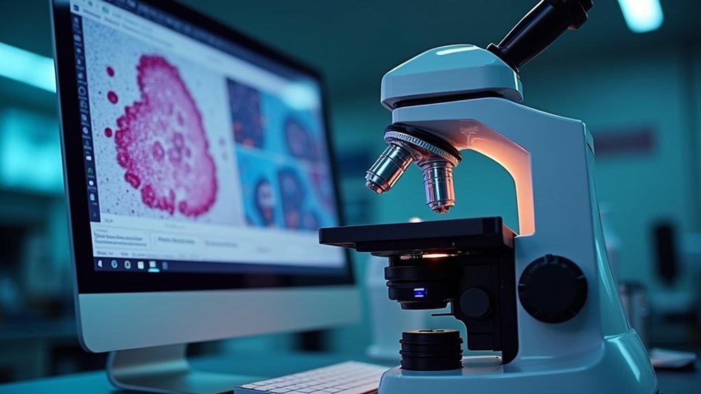
How to Improve Microscope Images?
You’ll improve microscope images by adjusting exposure settings, using proper illumination techniques, applying contrast enhancement filters, ensuring ideal focus, and utilizing noise reduction algorithms in your imaging software during acquisition and post-processing.
In Summary
When you’re selecting microscope imaging software, you’ll find each tool offers distinct advantages for your research needs. Whether you’re looking for commercial solutions like Leica’s LAS X and Zeiss ZEN, or prefer open-source options like ImageJ, there’s something for every budget and application. Don’t hesitate to try demos before committing—the right software can dramatically enhance your imaging capabilities and data analysis.


Leave a Reply