Author: SciCapture
-
5 Steps To Master Dark Field Lighting
Five critical techniques reveal dark field lighting’s secrets, transforming ordinary photos into striking images with dramatic textures.
-
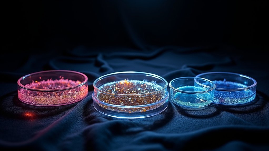
Top 3 Instruments For Stunning Darkfield Image Capture
Curious about creating breathtaking darkfield microscopy images? Discover the three essential instruments that transform invisible specimens into visual masterpieces.
-

10 Dark Field Condenser Tips for Sharp Images
From proper alignment to specimen preparation, these essential darkfield condenser adjustments will transform your microscopic images—but the most critical technique remains overlooked.
-
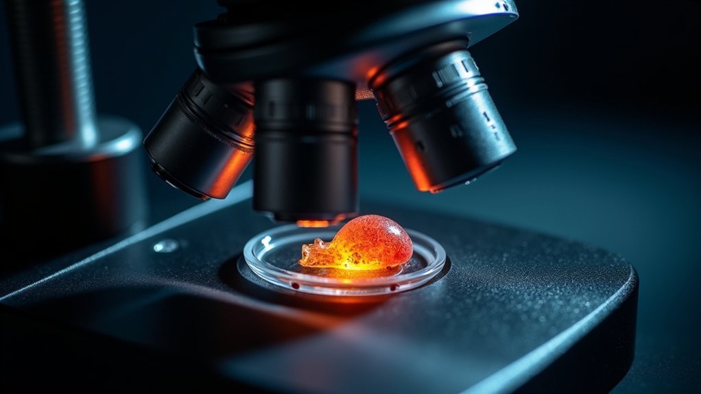
5 Pro Tricks For Stunning Darkfield Image Capture
Mastering darkfield microscopy transforms ordinary specimens into glowing treasures, but most photographers miss these essential lighting secrets.
-

7 Ways To Silence Noisy Stage Movements
Astonishing stage silence awaits performers with these 7 techniques that eliminate distracting noises during critical moments.
-

7 Ways To Test Stage Movement Precision
Journey through proven methods for measuring stage movement accuracy that will transform how you evaluate precision machinery.
-

Stage Positioning: Essential Tips for Precision Maintenance
Gain critical insights into stage positioning maintenance protocols that protect precision performance—discover why experts prioritize these overlooked procedures.
-

Installing Your Stage Controller: Step-by-Step Setup Guide
Wondering how to properly set up your stage controller for maximum performance? This guide reveals essential installation secrets that most users miss.
-

What Stops Stage Drift During Live Cell Imaging?
Tackle stage drift in live cell imaging with thermal systems, hardware autofocus, and specialized chambers—but which combination works best for your research?
-

Stage Position Memory: Capture Repeatable Lab Images
Achieving perfect microscope imaging requires stage position memory techniques that revolutionize laboratory documentation, but what’s the secret?