To capture clear acid-fast stains, prepare a thin, heat-fixed specimen on a clean slide using four loopfuls of material. Apply carbol fuchsin with steam, decolorize precisely, and counterstain with methylene blue. Use an oil immersion objective (100x) for ideal magnification, and adjust your camera settings to low ISO with moderate aperture. Reduce artifacts through proper microscope calibration and enhance images with digital stain normalization. These techniques transform even difficult samples into diagnostically valuable images.
Preparing Specimens for Acid Fast Staining
When preparing specimens for acid-fast staining, you’ll need to start with a properly prepared slide to guarantee reliable results.
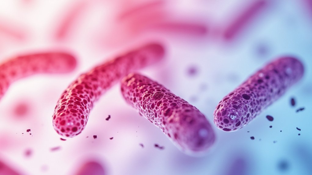
Begin by creating a thin film of microorganisms on a clean glass slide and allowing it to air dry. This prevents clumping and guarantees even visualization of acid-fast bacteria like Mycobacterium tuberculosis.
Next, heat fix your specimen by passing the slide through a flame three times. Let it cool before proceeding with staining procedures to avoid damaging the cells.
Use a wax pencil to label your slide and mark the specimen area with a circle. When you prepare the emulsion, add four loopfuls of the microbial sample to achieve the best coverage.
These careful preparation steps form the foundation for successful acid-fast staining results.
Essential Equipment and Materials for Clear Imaging
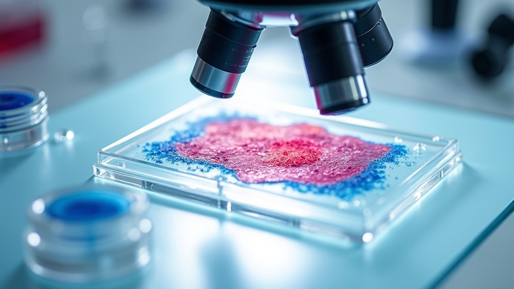
Your microscope’s quality directly affects your ability to identify acid-fast bacteria, with oil immersion objectives (100x) providing the resolution needed to see their distinctive red coloration against blue backgrounds.
You’ll achieve clearer images by adjusting the diaphragm and condenser to create ideal contrast, helping distinguish between acid-fast and non-acid-fast organisms.
Proper lighting is equally critical—too bright will wash out subtle staining differences, while insufficient illumination will obscure the characteristic red rods that indicate mycobacteria presence.
Microscope Quality Matters
The microscope serves as the foundation for successful acid-fast staining visualization. You’ll need a high-quality instrument equipped with an oil immersion objective at 100x magnification to properly identify acid-fast bacteria.
Don’t overlook the importance of clean glass slides and cover slips, as contamination can compromise your results regardless of staining methods used.
For maximum clarity, verify your microscope has appropriate lighting settings—either brightfield or phase contrast—to enhance the visibility of stained organisms.
Regular maintenance of your equipment isn’t optional; clean lenses prevent artifacts that could lead to misinterpretation.
If you’re documenting your findings, invest in a compatible digital camera system capable of capturing high-resolution images. This documentation proves invaluable for analysis, verification, and maintaining thorough laboratory records.
Proper Lighting Setup
Optimizing lighting conditions represents the critical cornerstone of successful acid-fast staining visualization. You’ll need proper lighting to distinguish acid-fast bacilli clearly—their characteristic red staining against blue backgrounds demands excellent illumination.
Ensure adequate ambient lighting in your lab to prevent shadows and glare on slides. Your microscope should feature a consistent light source—halogen or LED lights provide even illumination that enhances contrast between stained and unstained organisms.
When examining specimens, use the oil immersion objective for maximum resolution. Carefully adjust the diaphragm to control light intensity; too much brightness washes out details, while insufficient light obscures critical features.
For documentation purposes, consider attaching a digital camera to capture high-resolution images of your specimens, allowing for detailed analysis and record-keeping of your acid-fast staining results.
Optimizing Staining Technique for Maximum Contrast
To achieve exceptional clarity with acid-fast stains, proper staining technique becomes paramount. Your success depends on meticulous attention to each step of the process. Guarantee even coverage of carbol fuchsin while using a steaming technique to maintain moisture, facilitating better dye absorption into acid-fast bacteria’s waxy cell walls.
Meticulous technique and even carbol fuchsin application ensure optimal penetration through waxy bacterial walls for superior acid-fast staining results.
- Monitor decolorization carefully – over-processing with acid alcohol leads to false negatives, while proper timing preserves the bright red color of acid-fast organisms.
- Allow sufficient time for methylene blue counterstaining to develop proper contrast against the pink acid-fast bacteria.
- Examine slides using the oil immersion objective to maximize resolution and visibility of stained organisms.
Remember that contrast is your goal – the stark difference between bright red acid-fast bacteria and blue non-acid-fast organisms enables accurate identification.
Camera Setup and Microscope Calibration Tips
Capturing breathtaking acid-fast stain images requires proper equipment setup after you’ve mastered the staining technique itself. Secure your camera to the microscope, guaranteeing perfect alignment of both optical axes for peak image capture.
Set your camera to a low ISO to minimize noise and enhance clarity, especially at high magnifications. Choose a moderate aperture (f/8-f/11) to balance depth of field with image sharpness.
Before shooting, properly calibrate your microscope’s focus using the oil immersion objective—this guarantees bacteria appear crisp and well-defined.
To prevent blur from compromising your results, use a remote shutter release or timer function. This eliminates vibrations that could obscure fine details in your acid-fast staining.
These technical adjustments will transform good microscopy into exceptional scientific documentation.
Image Capture Methods for Acid Fast Bacilli
When you’re ready to document acid-fast bacilli, proper image capture techniques become essential for accurate representation and analysis. Utilize your high-resolution microscope with oil immersion objectives to reveal the distinctive red carbol fuchsin stain against the blue background of the stained slide.
- Timing matters – Capture images immediately after completing staining techniques to prevent fading of the carbol fuchsin stain, especially under fluorescent lighting.
- Focus precision – Adjust your microscope meticulously at each magnification level, as even slight focus issues can obscure the thin acid-fast bacilli.
- Documentation details – Always photo document your specimen with notes about the specific staining techniques used, magnification level, and equipment settings for reproducibility and future reference.
Post-Processing Techniques to Enhance Diagnostic Clarity
You’ll need to enhance background contrast in your captured acid-fast bacilli images by adjusting brightness and saturation levels to make the red-stained bacteria stand out against the blue counterstain.
To eliminate artifacts that might interfere with diagnosis, try using digital filtering tools that can remove dust particles, air bubbles, and staining precipitates without affecting the bacterial structures.
Digital stain normalization techniques can standardize color variations between different slides, ensuring consistent interpretation regardless of minor staining intensity differences.
Background Contrast Enhancement
To detect acid-fast bacteria effectively, digital post-processing techniques can markedly improve diagnostic clarity.
When identifying acid-fast bacteria, you’ll need to enhance images through background contrast enhancement to distinguish these organisms from surrounding tissues. The staining technique alone isn’t always sufficient—digital image processing can considerably improve clarity and diagnostic accuracy.
Here are three essential contrast enhancement methods:
- Apply histogram equalization to emphasize the bright red acid-fast organisms against blue/green backgrounds.
- Utilize edge enhancement algorithms to sharpen bacilli outlines, making them more distinguishable from non-acid-fast elements.
- Adjust brightness and contrast settings in your imaging software while maintaining the original specimen’s integrity.
Remember that while enhancing images, you must preserve the authentic diagnostic characteristics of the sample to guarantee reliable results.
Artifact Removal Strategies
Beyond contrast enhancement, artifact removal represents the next vital step in producing diagnostically valuable acid-fast stain images.
You’ll need high-quality imaging software to eliminate distractions that obscure acid-fast bacteria. Start by cropping your images to focus only on relevant areas, immediately improving clarity and diagnostic value.
Apply specialized filters to separate acid-fast bacilli from background debris—this helps considerably when quantifying acid-fast bacilli for accurate diagnosis.
Don’t overlook the importance of consistent calibration of your microscope before capturing images, as this prevents many artifacts from appearing in the first place.
For ideal results, adjust contrast levels systematically after removing artifacts. This two-step approach guarantees that what you’re analyzing is actual bacterial presence rather than image imperfections that could lead to misinterpretation.
Digital Stain Normalization
The effectiveness of digital stain normalization can greatly improve your ability to identify acid-fast bacilli in microscopic samples. By applying techniques like histogram equalization and contrast stretching, you’ll enhance visibility of stained organisms against background tissue.
Color deconvolution methods help differentiate between the bright red acid-fast bacteria and blue-stained background, considerably improving readability and interpretation accuracy.
For ideal results, consider these post-processing approaches:
- Apply Gaussian blur filters to reduce noise while preserving bacterial morphology for more accurate diagnosis.
- Implement image segmentation algorithms to isolate bacilli from surrounding tissue, making quantification possible.
- Utilize machine learning techniques on your normalized images for automated identification and classification of acid-fast organisms.
These digital enhancements transform difficult-to-interpret samples into clear, diagnostically valuable images.
Frequently Asked Questions
What Are the Steps of the Acid-Fast Stain in the Proper Sequence?
You’ll prepare a heat-fixed slide, flood with carbol fuchsin for 2-3 minutes, decolorize with acid alcohol, counterstain with methylene blue for 30 seconds, then observe under oil immersion microscope for red acid-fast bacteria.
What Is the Correct Order of Steps for the Acid-Fast Stain in Order to Differentiate Between an Acid-Fast and Non Acid-Fast Bacterium?
To differentiate bacteria types, you’ll first heat-fix your bacterial smear, apply carbol fuchsin, rinse with water, decolorize with acid alcohol, and finally counterstain with methylene blue. Acid-fast bacteria retain red; others appear blue.
What Is the Correct Order of Chemicals for the Acid-Fast Stain?
For acid-fast staining, you’ll need to apply carbol fuchsin as your primary stain first, then use acid alcohol for decolorization, and finally apply methylene blue as the counterstain. Don’t forget to rinse between steps.
What Is the Procedure for AFB?
You’ll prepare a heat-fixed smear, flood with carbol fuchsin and heat for 5 minutes, decolorize with acid alcohol, counterstain with methylene blue for 30 seconds, then examine under oil immersion for red acid-fast bacteria.
In Summary
You’ve now mastered the art of capturing clear acid fast stains. By properly preparing specimens, optimizing your staining technique, and calibrating your equipment, you’ll consistently produce high-quality images. Remember to use appropriate post-processing methods without altering diagnostic features. With these skills, you’re well-equipped to document and identify acid fast bacilli accurately, enhancing your diagnostic capabilities and contributing to effective patient care.

Leave a Reply