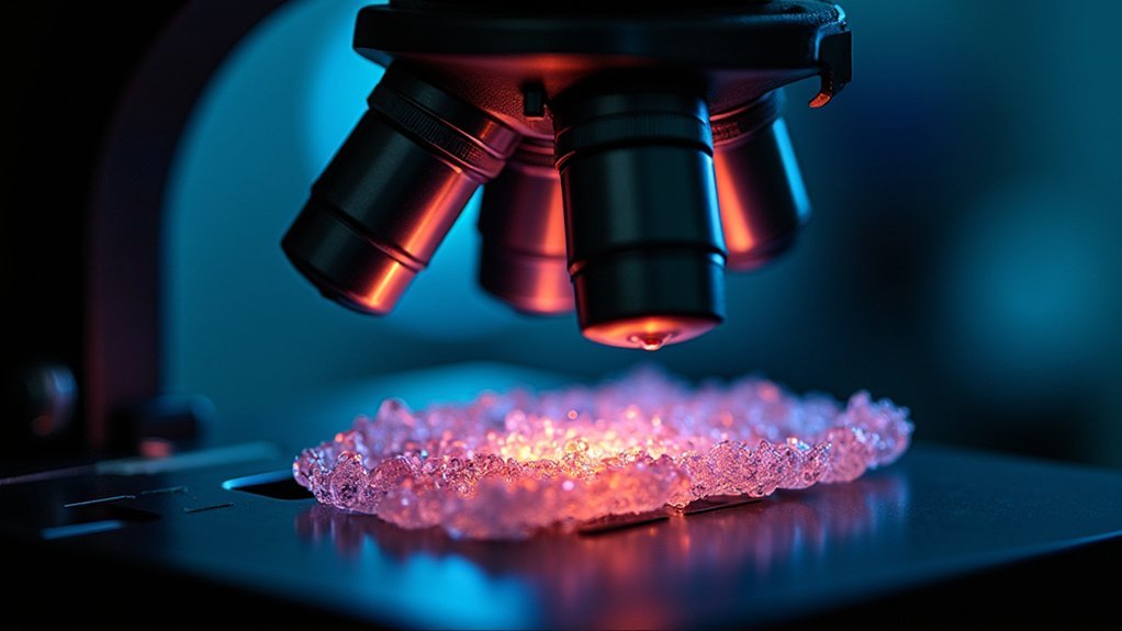
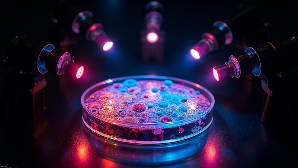
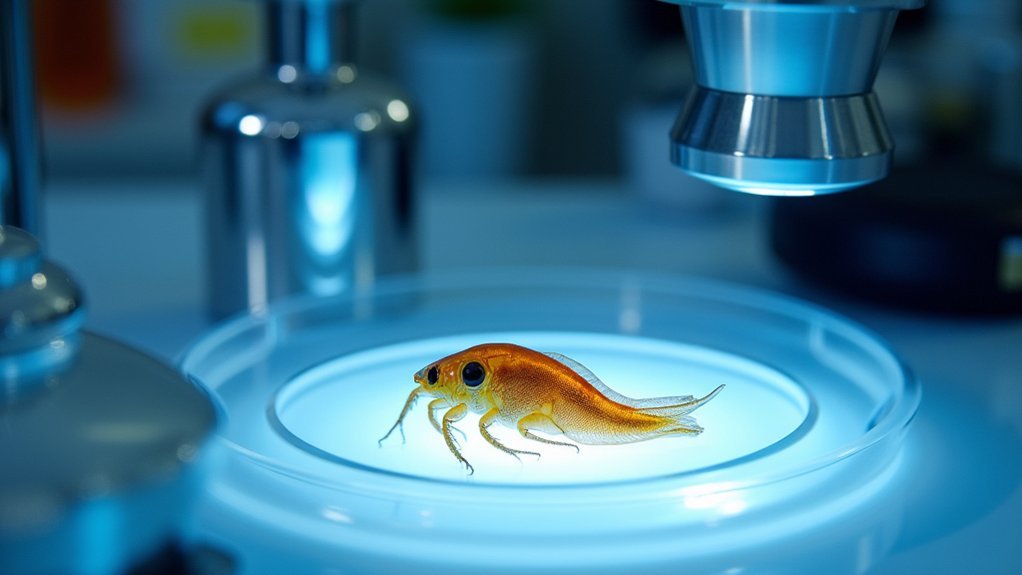
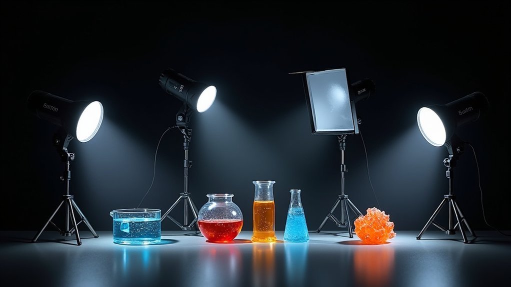
You’ll achieve superior scientific images with these five specialized strobe setups: Dual-Angle Microscope Systems that minimize shadows while highlighting textures, Ring Flash Configurations for shadow-free 3D visualization, Ultra-High Speed Strobes that freeze cellular motion at 1/1,000th second, Multi-Flash Arrays for creating detailed 3D reconstructions, and Diffused Single-Point Strobes that preserve delicate biological details. Each setup offers unique advantages for capturing specimens with remarkable clarity and precision you’ll discover below.
The Dual-Angle Microscope Strobe System for Specimen Detail

Precision is the hallmark of effective scientific photography, especially when documenting microscopic specimens. Unlike continuous lighting, the Dual-Angle Microscope Strobe System offers superior control when capturing detailed images of delicate or moving subjects.
You’ll find this setup particularly valuable because it illuminates specimens from two distinct angles, effectively minimizing shadows while highlighting textural details that might otherwise remain hidden.
By synchronizing high-speed strobe flashes with your microscope, you can freeze motion and capture incredibly sharp images without blur.
The real advantage comes in the adjustability—you can manipulate the strobe angles to create ideal contrast and depth in your photomicrographs.
Adjustable strobe angles deliver unparalleled control over contrast and depth, elevating your photomicrographs from mere documentation to visual excellence.
This precise control over exposure and aperture guarantees consistent image quality across varying conditions, making your scientific documentation more accurate and visually compelling for research publications.
Ring Flash Configuration for Shadow-Free Sample Imaging
While the Dual-Angle Microscope Strobe System excels at creating dimensional contrast, the Ring Flash Configuration represents a fundamentally different approach to scientific specimen illumination. You’ll achieve even, shadow-free lighting that’s essential when documenting intricate specimen details.
| Feature | Benefit | Application |
|---|---|---|
| Circular design | Surrounds subject with light | 3D structure visualization |
| Consistent color temperature | Accurate color representation | Specimen documentation |
| Close working distance | Enhanced detail capture | Microscopy work |
| Adjustable power output | Flexible exposure settings | Various sample types |
| Minimal post-processing | Time-efficient workflow | High-volume documentation |
The ring flash configuration saves you valuable time by reducing post-processing needs while maintaining compatibility with most camera systems. You’ll appreciate how this setup reveals fine details that might otherwise be obscured by shadows in conventional lighting arrangements.
Ultra-High Speed Strobes for Cell Movement Capture

When capturing the lightning-fast processes of cellular movement, ultra-high speed strobes become your essential tools for freezing action that the naked eye can’t perceive.
These specialized units achieve flash durations as brief as 1/1,000th of a second, eliminating motion blur even during rapid cellular activities like mitosis.
For ideal results, you’ll need powerful strobes exceeding 500 Ws that can effectively illuminate your microscopic subjects.
Don’t rely on just one light—position multiple strobes strategically around your specimen to minimize shadows and enhance vital details.
Look for models with built-in triggering systems that sync perfectly with high-speed cameras, ensuring you’ll capture the precise moment of cellular transformation.
This setup provides researchers with crystal-clear visual data that’s invaluable for analyzing complex biological processes in unprecedented detail.
Multi-Flash Array Setup for 3D Microscopic Reconstruction
The multi-flash array represents a revolutionary approach to microscopic imaging that transforms flat specimens into detailed three-dimensional models.
This setup typically requires positioning three to five strobe lights at different angles around your specimen to guarantee even illumination and shadow reduction.
When implementing this technique, make certain your strobes are synchronized with your camera’s electronic shutter for precise timing—critical for capturing clear, high-resolution images.
Unlike continuous lighting, strobes effectively freeze motion, which proves invaluable when examining dynamic subjects or using high magnification.
After capturing multiple perspectives, you’ll process these images through specialized software to generate thorough 3D reconstructions.
This technique greatly enhances visualization of complex microscopic structures, allowing you to examine details that would otherwise remain hidden in traditional flat imaging approaches.
Diffused Single-Point Strobes for Delicate Biological Samples

Three key advantages make diffused single-point strobes essential for capturing delicate biological specimens. First, they minimize harsh shadows and reflections, preserving vital details. Second, when positioned on light stands at a 45-degree angle, they enhance texture while maintaining soft illumination. Third, they’re easily modified with softboxes or diffusion materials to eliminate hotspots on translucent specimens.
| Power Setting | Application | Benefit |
|---|---|---|
| 1/16 power | Microscopic cells | Prevents overexposure |
| 1/8 power | Tissue samples | Balances detail/depth |
| 1/4 power | Larger specimens | Provides adequate fill |
| Variable | With color gels | Enhances specific features |
You’ll achieve ideal results using 1/16 to 1/4 power settings, controlling exposure while preserving intricate details. For advanced applications, integrate color gels to improve visibility of specific biological features.
Frequently Asked Questions
What Camera Settings for Strobe Photography?
You’ll want to set your camera to low ISO (100-200), fast shutter speed (1/200s+), appropriate aperture (f/2.8-f/16 depending on depth needed), and manual mode for full control with strobe photography.
Should I Get Strobe or Speedlight?
For general photography, choose strobes if you need power and consistency, speedlights if portability matters most. For scientific photography specifically, strobes are better due to their higher output, color accuracy, and modeling lights.
What Is the Best Shutter Speed for Strobes?
For strobes, you’ll want to use shutter speeds between 1/125 and 1/250 seconds. Don’t exceed your camera’s max sync speed or you’ll get banding in your images. Test your specific setup first.
In Summary
You’ve now explored five powerful strobe lighting techniques that’ll transform your scientific photography. Whether you’re capturing microscopic cell movement or creating 3D reconstructions, these setups will help you document specimens with unprecedented clarity and detail. Remember, the right lighting doesn’t just illuminate—it reveals scientific truth. Experiment with these configurations to find what works best for your specific research needs and imaging challenges.


Leave a Reply