To prevent fluorescence fading, reduce light intensity using neutral-density filters and optimize microscope settings before imaging. Use photostable fluorophores like Alexa Fluor or Atto dyes, and mount samples in appropriate antifade media for at least 2 hours. Keep exposure times short, employ image binning, and use transmitted light for focusing. Control temperature and oxygen levels during the entire process. These strategies will dramatically extend your fluorophore lifespan and improve quantitative analysis.
Understanding Photobleaching: The Science Behind Fluorescence Fading
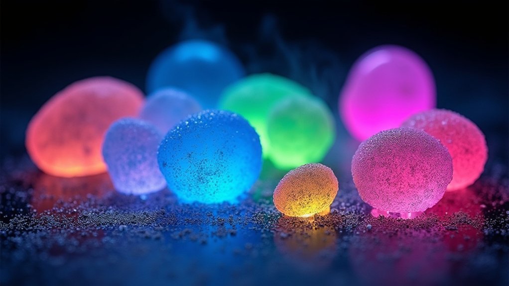
While fluorescence microscopy enables visualization of cellular structures with remarkable clarity, the phenomenon of photobleaching threatens the integrity of your observations. This chemical damage occurs when your fluorophores experience prolonged exposure to excitation light, permanently destroying their ability to fluoresce.
You’ll notice this fading effect most dramatically in samples with low-abundance targets, where even minor signal loss can compromise quantitative analysis. Photobleaching happens through covalent modifications when fluorophores react with each other or their environment.
Photobleaching’s fading effect can devastate low-abundance samples, compromising analysis when fluorophores undergo irreversible chemical reactions.
To preserve your valuable data, you should visualize samples immediately after fluorescent labeling, as signal diminishes rapidly following preparation. Using antifade mounting media can greatly extend fluorophore lifespan.
For precise analysis, consider creating photobleach curves to distinguish between normal photobleaching and other factors affecting your signal strength.
Selecting Photostable Fluorophores for Extended Imaging
How you choose your fluorophores can make or break your imaging experiments, especially when extended visualization is essential.
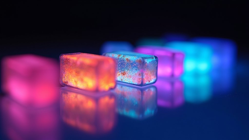
Modern dyes like Alexa Fluor, DyLight, and Atto offer superior photostability compared to traditional FITC and TRITC, which rapidly succumb to photobleaching in fluorescence experiments.
For ideal results when selecting fluorophores:
- Test your chosen dyes under your specific experimental conditions before committing to a full study
- Consider wavelength characteristics—some spectral regions naturally resist photobleaching better than others
- Implement dual labeling strategies with photostable dyes to maintain signal integrity throughout imaging
- Pair your photostable fluorophores with an appropriate antifade mounting medium to further extend fluorescence lifespan
These choices will dramatically improve your imaging duration while maintaining signal quality throughout your experiments.
Optimizing Light Exposure: Techniques to Reduce Excitation Damage

You’ll greatly extend your fluorophore’s lifespan by strategically reducing light intensity through neutral-density filters and optimized gain settings.
Time your exposures intelligently by using transmitted light for initial focusing, then switching to fluorescence only when absolutely necessary.
Implement image binning to decrease required exposure times, allowing you to capture clear images while minimizing the damaging effects of excitation light.
Minimizing Light Intensity
Excessive light exposure remains the primary culprit behind fluorescence fading, making strategic light management essential for preserving sample integrity. To prevent photobleaching, you’ll need to deliberately reduce the intensity of excitation light reaching your specimens.
Utilize neutral density filters to effectively decrease light levels without compromising detection sensitivity.
- Adjust your microscope’s gain settings instead of increasing light intensity for better visibility
- Implement image binning techniques to combine pixels, reducing necessary exposure while maintaining signal strength
- Focus on your region of interest using transmitted light before switching to fluorescence mode
- Use shorter exposure times during initial focusing phases to minimize cumulative photon damage
These minimizing light intensity strategies will notably extend your fluorophores’ lifespan while maintaining image quality for more reliable, reproducible results.
Smart Exposure Timing
Beyond reducing light intensity, intelligent timing of fluorophore exposure represents a powerful strategy for mitigating photobleaching. In fluorescence microscopy, you’ll get better results by using transmitted light for initial focusing rather than immediately exposing samples to excitation light.
When searching for regions of interest, implement less-than-ideal exposure times to minimize cumulative light damage.
You can further enhance your smart exposure timing by incorporating image binning techniques, which improve signal-to-noise ratios while reducing overall light exposure.
Don’t forget to utilize neutral density filters to control excitation intensity during your imaging sessions. Regularly adjusting your microscope’s gain settings helps strike the best balance between signal detection and minimal light usage.
These timing strategies work together to greatly extend your fluorophores’ lifespans and improve image quality throughout your experiments.
Antifade Mounting Media: Formulations and Best Practices
When preserving fluorescence signals during microscopy, your choice of mounting media can make the difference between crisp, long-lasting fluorescence and rapidly fading specimens.
Antifade mounting medium greatly reduces the loss of fluorescence during extended imaging sessions, particularly when you’re working with fixed cells.
For best results with your fluorescent samples:
- Test multiple antifade formulations as effectiveness varies depending on the specific fluorophore you’re using
- Consider p-phenylenediamine and glycerol-based solutions, which are particularly effective at minimizing photobleaching
- Allow samples to incubate in your chosen antifade medium for at least 2 hours (overnight is better)
- Remember that proper selection of antifade media can dramatically extend the visibility of your fluorescent signals
Sample Preparation Strategies to Preserve Fluorescence Intensity
Proper sample preparation forms the foundation of successful fluorescence microscopy, regardless of how sophisticated your imaging system might be. To maintain ideal fluorescence signal during imaging, process your samples immediately after labeling. If you can’t image right away, wrap samples in aluminum foil and store at 4°C in the dark.
| Strategy | Benefit | Impact |
|---|---|---|
| Immediate imaging | Maximum brightness | Stunning visuals |
| Use transmitted light first | Reduces photobleaching | Longer imaging sessions |
| Antifade mounting medium | Stabilizes fluorescence | Reliable quantification |
Choose appropriate fixation methods like paraformaldehyde or methanol to preserve cellular structure while maintaining epitope accessibility. These techniques guarantee your samples retain their fluorescence intensity throughout your imaging session, providing consistent and reliable results without premature fading.
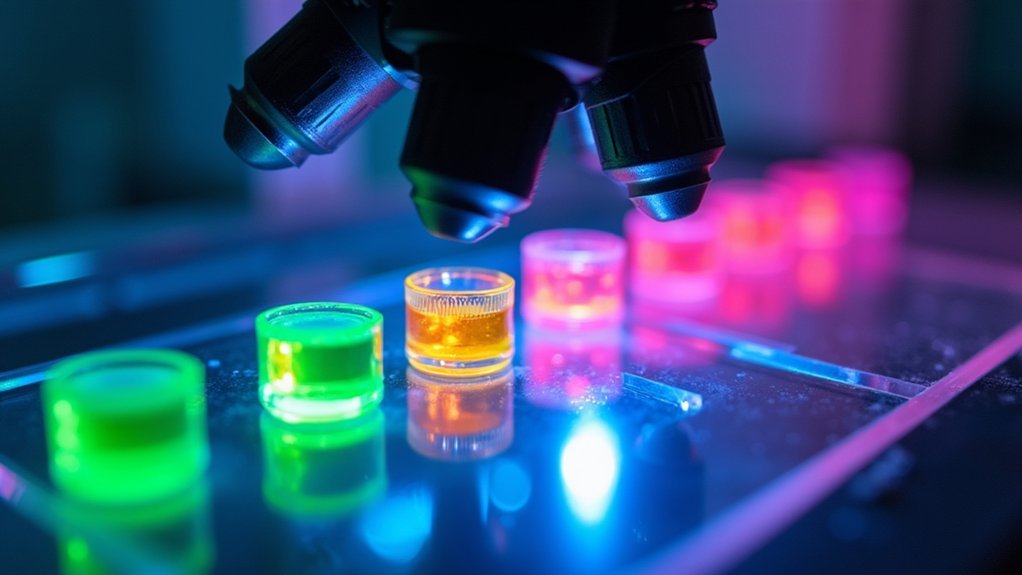
Advanced Imaging Settings to Minimize Photobleaching
Optimizing your microscope settings plays an essential role in preserving fluorescence signals throughout extended imaging sessions.
Strategic microscope optimization extends fluorophore lifespans, preserving critical signals during long imaging experiments.
You’ll see a significant improvement in your photobleach curve when you reduce light exposure through strategic adjustments. Implement antifade protection through your hardware settings rather than relying solely on mounting media.
- Switch to LED light sources instead of higher-intensity lamps to dramatically reduce photobleaching while maintaining adequate illumination
- Apply image binning techniques to improve signal-to-noise ratios without increasing exposure times
- Utilize neutral-density filters to attenuate excitation light, extending fluorophore lifespans
- Install and program microscope shutters to automatically block light between captures, preventing unnecessary sample exposure
Creating Photobleach Curves for Data Normalization
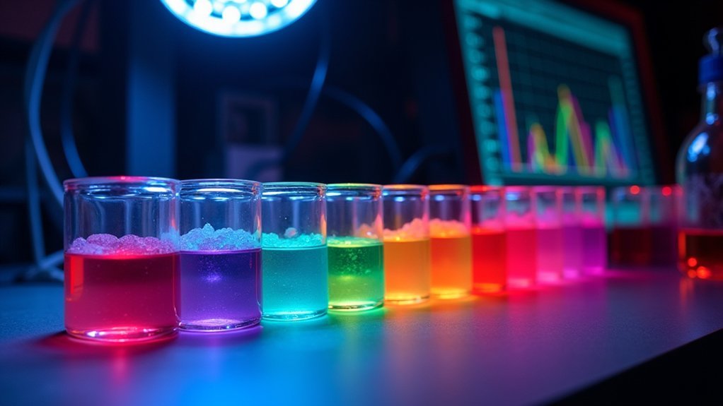
Photobleach curves provide essential benchmarks that help you separate genuine biological signals from imaging artifacts.
You’ll need to capture sequential images at fixed intervals and plot normalized fluorescence intensity against time to create these reference standards.
These curves then become powerful tools for data correction, allowing you to compensate for fluorescence loss in your experimental samples and extract more reliable quantitative measurements.
Why Curves Matter
While collecting fluorescence data might seem straightforward, understanding the inevitable decay in signal intensity over time is essential for accurate interpretation.
Photobleach curves provide a visual representation of this decay, allowing you to distinguish between true experimental effects and artifacts caused by fluorophore fading.
- Curves help you separate actual biological changes from normal fluorescence intensity loss
- They establish baseline photobleaching rates for normalizing future experimental data
- You’ll identify ideal imaging parameters that minimize unnecessary photobleaching
- They enable more accurate comparisons across different samples and experimental conditions
Plotting Step-by-Step
Now that we’ve established why photobleach curves matter, let’s build one from scratch. Start by capturing a time-series of fluorescence images at consistent intervals from the same region of interest. For each image, measure the average fluorescence intensity within your defined area.
Next, plot these intensity values against time on a graph to visualize how quickly your signal fades during Fluorescence Imaging. This creates your photobleach curve, revealing the decay rate specific to your experimental conditions.
To normalize your experimental data, divide each measurement by the corresponding value on your photobleach curve. This essential step separates genuine biological signals from artifacts caused by photobleaching.
A well-constructed photobleach curve isn’t just for data correction—it helps you optimize exposure times and select more photostable fluorophores for future imaging experiments.
Environmental Factors Affecting Fluorescence Stability
When you’re working with fluorescent specimens, understanding the environmental conditions around your samples can make the difference between crisp, stable signals and rapidly fading fluorescence.
Your imaging environment directly impacts how long your fluorophores will maintain their intensity.
- Temperature control is essential—higher temperatures accelerate photobleaching reactions and diminish signal longevity.
- Light exposure should be minimized before and during imaging as ambient light can trigger premature photobleaching.
- Oxygen levels near your sample can cause oxidative damage to fluorophores, resulting in signal degradation.
- Proper mounting with antifade reagents provides protection against environmental factors, creating a shield around your fluorophores.
Frequently Asked Questions
How Do You Prevent Fluorescence?
To prevent fluorescence, you’ll need to use antifade mounting media, minimize exposure time, reduce light intensity with neutral density filters, and choose photostable dyes like Alexa Fluor for your samples.
How Can Photobleaching Be Prevented?
To prevent photobleaching, you’ll want to use resistant fluorophores like Alexa Fluor, implement neutral-density filters, maintain consistent imaging settings, apply anti-fade mounting media, and employ FRAP for intensity normalization when needed.
How to Reduce Background Fluorescence?
To reduce background fluorescence, use blocking agents, optimize washing steps, select non-overlapping fluorophores, utilize clean coverslips, and implement proper controls. You’ll achieve clearer images with these targeted approaches to minimize non-specific signals.
Does Fluorescence Fade?
Yes, fluorescence does fade. It’s called photobleaching, which happens when your fluorophores lose their ability to fluoresce after prolonged light exposure. You’ll see this as diminishing signal intensity during imaging.
In Summary
You’ll find that preventing fluorescence fading isn’t complex once you’ve mastered these fundamentals. By choosing stable fluorophores, optimizing light exposure, using quality antifade media, and adjusting your imaging settings, you’ll capture clearer images with minimal photobleaching. Remember, it’s the combination of these techniques—not just one solution—that will dramatically extend your fluorescence signal and improve your data quality.


Leave a Reply