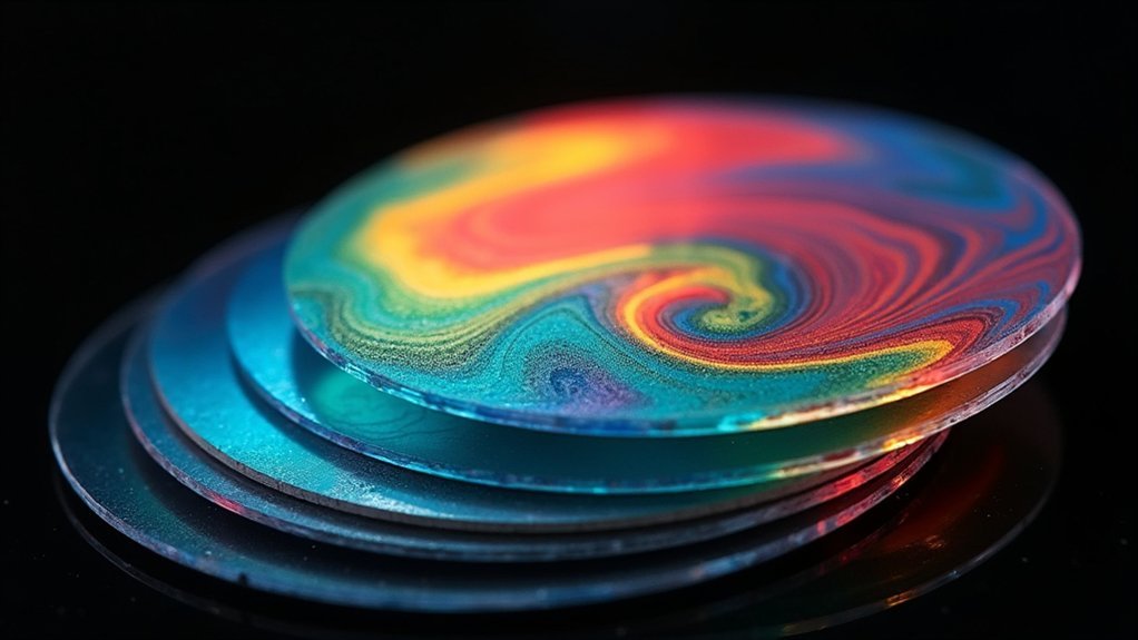
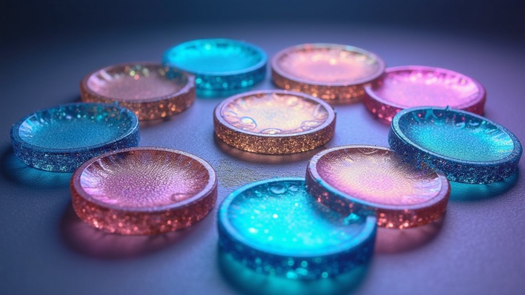
For stunning polarized microscopy images, you’ll need first-order red (λ) plates for vibrant magenta backgrounds, quarter-wave (λ/4) plates for circular polarization, and half-wave (λ/2) plates for rotation control. Don’t forget full-wave plates and quartz wedge compensators for crystalline analysis. Berek compensators offer precise measurements, while polymer films provide cost-effective alternatives. Achromatic wave plates eliminate chromatic dispersion across wavelengths. The Michel-Lévy chart helps interpret those beautiful interference colors you’ll soon master.
First Order Red (λ) Retardation Plates: Enhancing Contrast in Weakly Birefringent Specimens

Microscopists rely on first order red (λ) retardation plates as essential tools for visualizing subtle structures in polarized light microscopy. When you’re examining specimens with minimal birefringence, these plates introduce a fixed optical path difference between 530-560nm, transforming barely visible details into vivid displays.
The retardation plates create approximately one wavelength (550nm) of relative retardation, producing a distinctive magenta background as green wavelengths are removed. You’ll find this particularly useful when determining the optical sign of birefringent materials—positive when the extraordinary wavefront travels slower, negative when the ordinary wavefront lags.
For best results, position these plates at a 45-degree angle to your polarizer and analyzer. They’re especially effective for biological specimens like cell walls and starch granules, revealing interference colors and details invisible in conventional microscopy.
Quarter Wave (λ/4) Plates: Converting Linear to Circular Polarization
Quarter wave plates transform linearly polarized light into circular polarization through a precise 90-degree phase shift between orthogonal components.
You’ll find these optical elements constructed from birefringent materials like quartz, mica, or polymer films with carefully controlled thickness to match specific wavelengths.
In polarized light microscopy, λ/4 plates help you visualize specimens with directional properties, enabling techniques like circular dichroism imaging and reducing unwanted glare from reflective samples.
Optical Properties Overview
The remarkable ability of quarter wave (λ/4) plates to manipulate polarized light stands as a cornerstone in optical engineering. These special optical elements introduce a precise 90-degree phase shift between the fast and slow axes of polarized light passing through them.
When you align a λ/4 plate at 45 degrees to incoming linearly polarized light, you’ll witness a fascinating transformation: the light emerges circularly polarized. This occurs because the birefringent materials in the plate cause one component of light to travel faster than the other, creating the necessary retardation effect.
You’ll find these plates essential in numerous applications, from optical isolators to polarization-sensitive detection systems. Their performance depends critically on wavelength matching, as the precise retardation effect must be calibrated to the specific light source you’re working with.
Materials And Construction
Building on our understanding of optical properties, let’s examine how λ/4 plates achieve their remarkable effects through careful material selection and precise construction. When a retardation plate is inserted into your optical path, it creates a controlled optical path difference between perpendicular polarization components.
True zero order plates use ultra-thin birefringent materials like quartz, providing excellent stability against temperature and wavelength fluctuations. These plates work by exploiting the material’s different refractive index values along fast and slow axes.
For broader wavelength applications, you’ll find achromatic quarter wave plates invaluable, as they combine two birefringent materials to maintain consistent 90-degree phase shifts across multiple wavelengths.
You can further enhance polarization control by pairing λ/4 plates with λ/2 plates, creating custom configurations for your specific imaging needs.
Applications In Microscopy
When exploring the powerful domain of optical microscopy, λ/4 plates emerge as indispensable tools for transforming polarization states and enhancing specimen visibility.
You’ll find these quarter wave plates particularly valuable when examining birefringent materials where polarized light interactions reveal essential structural details.
- Position your λ/4 plate at precisely 45° to incoming linearly polarized light to generate circular polarization, eliminating unwanted interference colors in highly birefringent samples.
- Create stunning contrast in anisotropic specimens by controlling light transmission through the sample.
- Stack λ/4 plates with λ/2 plates to achieve custom retardation values for specialized imaging requirements.
- Toggle between linear and circular polarization states to highlight different aspects of your specimen’s structure.
Proper alignment is vital—even slight misalignment will compromise your polarization control and diminish image quality in polarized light microscopy applications.
Half Wave (λ/2) Plates: Rotating the Plane of Polarization
When selecting materials for half wave plates, you’ll need to take into account your application’s specific wavelength requirements and temperature sensitivity.
For precision rotation applications, proper mounting and alignment of your λ/2 plate will greatly impact polarization accuracy and stability.
You can achieve ideal performance by pairing high-quality crystalline quartz plates with precision rotation mounts that offer fine angular adjustment capabilities.
Precision Rotation Applications
Three key rotation applications make half wave (λ/2) plates essential tools in precision optics.
When you position these plates at precise angles relative to incoming polarized light, you’ll achieve exact rotation control. Remember that the output polarization rotates at twice the physical angle you set the plate at, giving you incredible precision when manipulating light paths.
- Optical isolators – Prevent unwanted back-reflections by rotating polarization states
- Polarized light microscopy – Enhance contrast in birefringent samples by fine-tuning polarization angles
- Laser beam control – Adjust beam polarization to match specific optical component requirements
- Quantum optics – Prepare photons in specific polarization states for quantum information experiments
The fast axis orientation determines how effectively you’ll rotate the incoming light, making proper alignment critical when you need exact angle of rotation in high-precision applications.
Material Selection Guide
Every effective half wave plate depends critically on your material selection based on the target wavelength range. When choosing materials for your λ/2 plate, consider birefringent options like quartz or magnesium fluoride, which perform best across specific spectral ranges.
Remember that retardation is wavelength-dependent, so matching your material to your light source is essential.
Pay close attention to thickness—typically around 0.5 mm for standard applications—as this parameter determines the quality of your polarized light transformation. The right thickness guarantees your plate achieves the precise 180-degree phase shift without introducing unwanted wavelength variations.
For best results, select plates with anti-reflective coatings to minimize light loss and maximize transmission efficiency. Proper mounting will further enhance your polarization control capabilities.
Full Wave Plates: Revealing Optical Sign in Crystalline Materials

Full wave plates serve as critical tools in polarized light microscopy by introducing a precise 180-degree phase shift between orthogonal light components.
When you position these plates at a 45-degree angle to your polarizer and analyzer, they’ll enhance contrast and reveal the optical sign of birefringent crystalline materials.
- Distinguish between positive and negative birefringence based on how light travels through your specimen
- Create distinctive interference colors that highlight crystalline structures
- Generate conoscopic interference patterns when used with a Bertrand lens
- Enhance visibility of anisotropic features that would otherwise remain hidden
Quartz Wedge Compensators: Measuring Variable Retardation Effects
Sliding a quartz wedge compensator into your polarizing microscope’s slot introduces a powerful capability for measuring precise retardation effects.
The wedge’s ingenious thickness gradient creates continuous variation in optical path difference, allowing you to observe dynamic interference patterns under polarized light.
You’ll find these compensators exceptionally versatile, measuring retardation from just nanometers to multiple wavelengths.
As you rotate the wedge, watch for changes in color and brightness that directly correspond to varying levels of birefringence in your specimen. This visual feedback provides significant insights into the material’s optical properties.
For complex crystalline structures, you’ll appreciate how the wedge helps identify optical signs and birefringence characteristics with remarkable accuracy.
The ability to fine-tune retardation makes quartz wedges indispensable for detailed analysis of anisotropic materials.
Achromatic Wave Plates: Maintaining Consistent Retardation Across Wavelengths

Unlike standard retardation plates, achromatic wave plates solve one of polarized microscopy’s most persistent challenges by maintaining consistent retardation across multiple wavelengths.
When you’re working with varied light sources, these specialized plates guarantee your polarized images remain accurate and vibrant.
The innovative design combines two birefringent materials with precise alignment to combat chromatic dispersion, giving you:
- Consistent phase shifts regardless of the wavelength you’re using
- Enhanced image quality by eliminating chromatic errors in your observations
- Superior performance in spectroscopy applications where wavelength variation occurs
- More accurate polarization control for demanding scientific and industrial applications
You’ll find achromatic wave plates indispensable when your work demands high precision across the light spectrum, especially in polarized light microscopy and advanced imaging techniques.
Berek Compensators: Precise Measurement of Birefringence
Complementing achromatic wave plates in polarized microscopy are Berek compensators, specialized optical instruments that excel at measuring birefringence with exceptional precision.
You’ll find these devices particularly valuable when you need to determine exact optical path differences in your specimens.
The compensator’s dual rotating plates allow you to finely adjust retardation values, producing measurable changes in color or brightness as you rotate your sample.
Fine-tune your observations with dual rotating plates that reveal subtle optical changes as your specimen moves.
This enables quantitative analysis that’s difficult to achieve with standard retardation plates.
When studying crystalline materials or complex biological structures, you can use the resulting interference patterns to determine the sign of birefringence, enhancing your understanding of the material’s optical properties.
The precise measurements provided by Berek compensators are essential for proper characterization of birefringent specimens in advanced microscopy applications.
Michel-Lévy Chart: Interpreting Interference Colors for Material Identification

When examining birefringent specimens under polarized light, the Michel-Lévy chart serves as an essential analytical tool that transforms colorful optical phenomena into quantifiable data.
You’ll find this chart particularly valuable for determining both the magnitude and sign of birefringence in your samples.
- First Order interference colors (0-550nm) provide the most diagnostic information for material identification, with subtle color changes indicating specific optical properties.
- The Michel-Lévy chart enables you to calculate retardation using the formula R = t(n₂ – n₁), where specimen thickness is typically standardized at 30 microns.
- Higher order colors help determine the sign of birefringence – positive in first/third quadrants, negative in second/fourth.
- Your accuracy depends critically on precise thickness measurements, ensuring reliable identification across various mineral and biological specimens.
Polymer Retardation Films: Cost-Effective Alternatives for Polarized Imaging
While the Michel-Lévy chart helps you interpret crystalline optical properties, you’ll find polymer retardation films offer practical advantages for your polarized imaging needs.
These flexible alternatives to traditional mineral thin sections deliver consistent optical performance without the high cost.
You’ll appreciate how these films can achieve specific retardation values comparable to zero-order waveplates while being lightweight and easily mountable in portable systems.
Their versatility makes them ideal for both LCD applications and specialized polarized light imaging setups.
The large-scale manufacturing process keeps costs down without sacrificing quality—a significant benefit when you’re working with budget constraints.
If you’re looking to enhance your imaging capabilities without investing in expensive crystalline components, these polymer films provide an accessible entry point into advanced polarized light techniques.
Super-Achromatic Wave Plates: Eliminating Chromatic Dispersion in High-Resolution Imaging

Unlike standard retardation plates, super-achromatic wave plates provide exceptional consistency across the entire visible spectrum, eliminating the wavelength-dependent variations that typically compromise image quality.
You’ll achieve superior polarized light imaging by incorporating these specialized optical components into your microscopy setup.
These advanced wave plates effectively combat chromatic dispersion through:
- Multi-material construction using quartz, magnesium fluoride, and sapphire
- Maintenance of constant retardation regardless of wavelength fluctuations
- Significant reduction in optical aberrations for enhanced structural clarity
- Precise phase control that enables accurate birefringent analysis
When you’re conducting detailed research in mineralogy, biology, or materials science, super-achromatic wave plates deliver the resolution and accuracy required for critical observations.
Their ability to maintain performance across varied spectral conditions makes them essential for researchers needing reliable, high-resolution polarized imaging results.
Frequently Asked Questions
What Are Retardation Plates?
Retardation plates are optical components you’ll use to introduce phase shifts between polarized light waves. They’re made of birefringent materials like quartz and help you visualize and analyze weakly birefringent specimens in microscopy.
What Is the Retardation Plate on a Microscope?
A retardation plate on your microscope introduces a wavelength delay (~550nm) when placed at a 45° angle. It’ll enhance contrast in weakly birefringent specimens and help you determine their optical sign under polarized light.
What Does a Half-Wave Plate Do to Linearly Polarized Light?
A half-wave plate rotates your linearly polarized light by twice the angle between the plate’s fast axis and the light’s polarization. When set at 45°, it’ll completely flip your polarization direction by 90°.
When Polarized Light Is Needed for Imaging a Microscope?
You’ll need polarized light when examining birefringent specimens, visualizing internal stresses, studying crystalline structures, viewing anisotropic materials, or enhancing contrast in transparent samples that wouldn’t be visible in standard brightfield microscopy.
In Summary
You’ve now explored the 10 essential retardation plates for polarized microscopy. From first-order red plates to super-achromatic options, each serves a unique purpose in revealing structural details invisible to conventional imaging. Whether you’re examining crystals, biological specimens, or material stress patterns, you’ll achieve stunning results by selecting the right plate for your specific application. Don’t hesitate to experiment with these powerful optical tools.


Leave a Reply