
Polarized image resolution in photomicrography depends on multiple factors including your objective’s numerical aperture, precise 90° alignment of polarizing filters, sensor specifications, and sample preparation quality. High-quality objectives with appropriate NA values capture more detail, while proper polarizer-analyzer alignment guarantees only specimen-modified light reaches the camera. Your light source quality, environmental stability, and digital sensor technology (CMOS, CCD, sCMOS) also greatly impact final image clarity. Understanding these elements will transform your polarized microscopy results.
Optical Components and Their Impact on Polarized Light
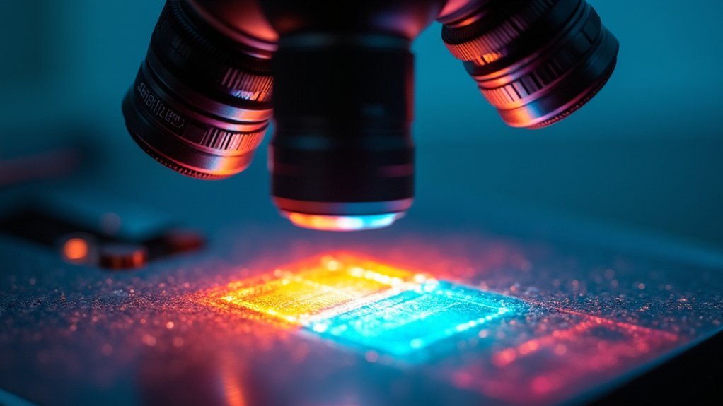
When setting up a polarized light microscope for photomicrography, you’ll need to confirm all optical components work in perfect harmony to achieve maximum resolution.
The precise 90° alignment of polarizing filters is critical—this crossed-polarizer configuration guarantees that only light modified by your specimen reaches the camera sensor.
Precise 90° filter alignment ensures only specimen-modified light reaches your sensor, revealing true birefringent properties.
Your choice of objectives greatly affects image quality. Select high-quality lenses with appropriate numerical apertures to capture more light and reveal finer details of specimens with varying refractive indices.
Don’t overlook compensators like wave plates, which enhance contrast and color in birefringent materials.
Remember that even minor misalignments in the optical train will compromise resolution.
Regular calibration of all components is essential, as proper alignment guarantees crisp, clear images that accurately represent the specimen’s birefringent properties.
Numerical Aperture and Resolution Limits in Polarization Microscopy
Resolution power in polarized light microscopy hinges directly on your objective’s numerical aperture (NA). The formula NA = n × sin(θ) tells you that higher refractive index media and wider collection angles greatly improve your resolution capability.
You’ll achieve finer detail as NA increases, with resolution limits approximated by λ/(2NA).
When working with birefringent specimens, your high-NA objectives not only capture more detail but also collect considerably more light, enhancing image brightness and contrast. This becomes particularly valuable when you incorporate compensators and polarizers in your optical setup.
Remember that shorter wavelengths further improve resolution—a principle you can leverage when photographing delicate structures in polarized light microscopy. The difference between standard and high-NA objectives becomes immediately apparent in the crisp details of your photomicrographs.
Polarizer-Analyzer Alignment Techniques for Optimal Clarity
To achieve the best resolution in polarized photomicrography, you’ll need to master extinction angle precision by carefully rotating the polarizer and analyzer to their 90-degree crossed position.
Cross-polarization adjustment methods involve incremental fine-tuning of both optical elements while observing the darkfield condition, ensuring only birefringent light from your specimen reaches the camera.
You can further enhance contrast by implementing compensators, maintaining clean optical surfaces, and utilizing the rotating stage to align your specimen’s birefringent structures with the polarization axes.
Extinction Angle Precision
Achieving superior image resolution in polarized light microscopy depends critically on precise extinction angle determination. When your birefringent specimens are oriented at this specific angle, they’ll appear black against a dark background, dramatically enhancing contrast and detail in your photomicrographs.
You’ll need to verify proper polarizer-analyzer alignment at exactly 90° to achieve the ideal “dark position.” By utilizing a rotating stage, you can systematically adjust your specimen’s orientation until you identify the precise extinction angle—a value that varies between different materials and provides valuable information about their optical properties.
Don’t overlook the importance of consistent calibration of your microscope components. This maintenance guarantees your polarizer-analyzer alignment remains accurate across multiple observations, preserving the resolution quality that makes polarized light microscopy such a powerful analytical tool.
Cross-Polarization Adjustment Methods
While many factors affect image quality in polarized photomicrography, precise cross-polarization alignment stands as the foundation for exceptional clarity.
When polarized light passes through your specimen, ideal resolution depends on your technique:
- Position your polarizer and analyzer at exactly 90° to achieve the “dark position,” ensuring only light altered by the specimen’s birefringence reaches your camera.
- Use the rotating stage to align your specimen’s optical properties with the polarizers, maximizing contrast based on refractive index differences.
- Incorporate compensators or wave plates to introduce specific phase shifts that reveal subtle structural details otherwise invisible.
- Regularly check calibration and observe your specimen at different rotation angles to capture varying interference colors and enhance structural resolution.
Contrast Enhancement Techniques
Beyond basic cross-polarization, mastering precise alignment techniques between polarizer and analyzer will dramatically elevate your image quality.
Verify your polarizing elements are positioned at perfect 90° angles to achieve the darkest possible field, which creates ideal contrast enhancement in your specimens.
Rotate your sample stage methodically to find the orientation that best reveals birefringent structures in your specimen. You’ll notice significant differences in clarity as you adjust.
Don’t overlook the value of compensators and wave plates—these tools alter light phase to highlight specific features when using a light microscope.
Keep your optical path immaculately clean; even minor dust or scratches on polarizing filters can introduce artifacts that compromise resolution.
Finally, experiment with specimen thickness, as this directly influences interference colors and ultimately enhances interpretable detail in your photomicrographs.
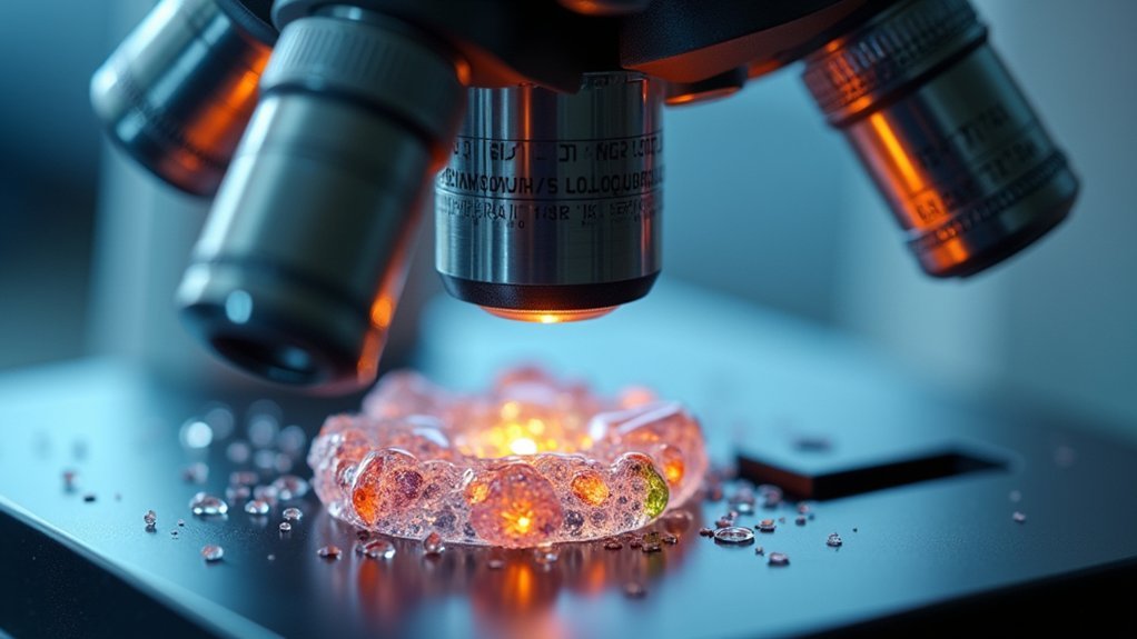
Sample Preparation Methods for Enhanced Birefringence Imaging

Since the quality of polarized images depends heavily on how specimens are prepared, mastering proper sample preparation techniques is essential for successful birefringence imaging.
Your attention to detail during this critical stage will greatly impact the visibility of birefringent materials and minimize optical aberrations.
For ideal results, implement these key preparation methods:
- Guarantee consistent sectioning and mounting to maintain even specimen thickness, reducing light scattering.
- Apply matching refractive index immersion oils to improve light transmission through your samples.
- Control drying conditions to promote proper crystal formation with well-defined structures.
- Utilize specialized staining agents and compensators to enhance contrast in birefringent structures.
Light Source Quality and Its Effect on Image Resolution
The light source you select dramatically impacts resolution quality in polarized photomicrography, determining your ability to capture fine structural details.
When working with polarized light, brightness and uniformity enhance contrast, revealing subtle features that might otherwise remain hidden.
LED illumination provides stable, vibration-free light that won’t introduce unwanted artifacts through heat distortion.
You’ll achieve better image resolution by selecting light sources with wavelengths that optimize birefringent properties in your specimens, enhancing differentiation based on refractive indices.
Be vigilant about light intensity—too much creates glare that obscures detail, while insufficient illumination reduces clarity.
Always verify your light source is properly calibrated and aligned with polarizing filters to maximize polarized light reaching the specimen.
This careful optimization directly translates to higher resolution imaging and more informative results.
Digital Sensor Technology in Polarized Photomicrography

Modern digital sensors transform your polarized photomicrography by offering resolution capabilities between 12-50 megapixels, which directly affects how much microscopic detail you’ll capture.
You’ll notice that pixel size fundamentally determines your ability to resolve micron-level structures, with smaller pixels enabling finer detail observation but potentially introducing noise in suboptimal lighting.
When selecting equipment for polarized microscopy, you should consider sensors with advanced technologies like back-illuminated designs that enhance light-gathering efficiency at the microscopic scale.
Sensor Resolution Impact
Digital sensor technology fundamentally transforms how polarized light is captured and represented in photomicrography, with resolution capabilities directly affecting image quality.
When you’re working with birefringent specimens, your sensor’s specifications determine how accurately you’ll capture interference colors and fine structural details.
Four vital factors affecting your polarized imaging quality:
- Pixel density – Higher counts enable you to resolve finer structures in your specimens, important for detailed digital imaging of crystalline materials.
- Sensor size – Larger sensors collect more light, reducing noise in low-light polarized microscopy conditions.
- Dynamic range – Wider ranges capture subtle variations in interference colors and contrast.
- Bit depth – Higher values preserve more tonal gradations, fundamental for accurate representation of polarization effects.
Micron-Level Detail Capture
When examining specimens at the cellular or crystalline level, your ability to resolve micron-scale features depends considerably on how effectively your digital sensor converts polarized light into meaningful data. Modern microscope systems utilize advanced digital sensors that capture incredibly fine details beyond what the human eye can discern.
| Sensor Type | Resolution Capability | Performance in Polarized Light |
|---|---|---|
| CMOS | 0.5-2 microns | Good contrast, moderate sensitivity |
| BSI CMOS | 0.2-0.5 microns | Excellent in low light, high sensitivity |
| CCD | 0.5-1 micron | Superior uniformity, moderate speed |
| sCMOS | 0.1-0.3 microns | High dynamic range, fast acquisition |
| EM-CCD | 0.2-0.5 microns | Exceptional in extremely low light |
Larger sensors with higher pixel density will enhance your resolution capabilities, allowing you to distinguish structural elements that are mere microns apart while maintaining clarity in polarized light conditions.
Post-Processing Techniques for Polarized Microscope Images
Although capturing high-quality polarized microscope images requires precise optical setup, the real enhancement often occurs during post-processing.
While the microscope captures the initial image, post-processing techniques truly reveal polarized light’s hidden details.
When working with polarized light microscope images, you’ll need several key techniques to maximize detail and accuracy:
- Brightness and contrast adjustments – Enhance visibility of birefringent structures and make interference colors more discernible.
- Image stacking – Combine multiple focal plane images to improve depth of field and overall clarity.
- Color balancing – Guarantee accurate representation of interference colors to reflect true sample characteristics.
- Specialized software applications – Use tools like Adobe Photoshop or dedicated analysis software to apply filters, reduce noise, and sharpen details.
Creating composite images also allows for side-by-side comparison of different samples, making your analysis more thorough and informative.
Environmental Factors Affecting Polarized Image Quality

While post-processing can enhance polarized microscope images, even the most sophisticated techniques can’t compensate for environmental issues present during image capture.
You’ll need to control lighting conditions meticulously to minimize shadows and glare that degrade image quality.
Temperature fluctuations directly impact the refractive indices of your specimens, altering their birefringence and compromising resolution.
Similarly, humidity affects optical properties, particularly in biological samples, potentially reducing clarity and contrast.
Don’t overlook airborne particles and dust—these light-scattering culprits can appreciably diminish image quality.
The stability of the microscope is equally vital; even minor vibrations can misalign your polarizers and analyzers, resulting in blurred images.
Frequently Asked Questions
How Is Polarization Determined?
Polarization is determined by your filter orientation, typically set at 90° angles. You’ll find it’s also affected by your specimen’s material properties, light wavelength, sample thickness, and proper microscope calibration for ideal results.
What Is the Principle of Polarized Optical Microscopy?
Polarized optical microscopy uses crossed polarizers to analyze birefringent samples. You’ll see your specimen reveal structural details through interference patterns when light passes through it, highlighting molecular arrangements invisible in standard microscopy.
What Mechanism Is Used to Produce Images by Polarizing a Microscope?
You’ll produce polarized microscope images when light passes through a polarizer, interacts with birefringent samples, and reaches the analyzer. The analyzer only transmits light modified by the sample, creating high-contrast images against a dark background.
What Is the Concept of Polarization in Microscopic?
In microscopy, polarization filters light waves to oscillate in specific directions. You’ll see this enhances contrast in transparent specimens by allowing only light that’s changed orientation to pass through, revealing structures otherwise invisible.
In Summary
You’ll find that polarized image resolution in photomicrography depends on multiple interconnected factors. Your optical components, numerical aperture, and precise polarizer-analyzer alignment all play essential roles. Don’t overlook sample preparation and light source quality. Your digital sensor technology greatly impacts final image clarity, while thoughtful post-processing can enhance detail. Remember that environmental factors like vibration and temperature fluctuations will affect your results, so control these variables for ideal resolution.


Leave a Reply