
To achieve success in scientific research imaging, you'll need to follow established standards and protocols. Start by implementing DICOM standards for data exchange, capture images at ideal resolutions (300-600 ppi), and use proper color profiles like AdobeRGB. You'll want to maintain thorough documentation, conduct regular calibrations, and guarantee proper data security through encryption and access controls. There's much more to mastering digital imaging standards that can elevate your research quality.
Core Standards for Digital Image Acquisition
While digital imaging revolutionizes scientific research, establishing core standards for image acquisition remains essential for data integrity and interoperability. You'll find that following DICOM standards guarantees seamless data exchange across different imaging platforms and modalities.
Standardized digital imaging protocols ensure reliable data sharing and analysis across the scientific community, with DICOM leading the way forward.
When conducting ocular imaging studies, you must adhere to guidelines set by the AAO and ARVO to maintain quality and support AI applications. You'll want to implement lossless compression techniques for your pixel data to preserve image quality while enhancing storage efficiency.
For peak research outcomes, verify your imaging devices and PACS systems can provide machine-readable, discrete data from user-selected reports.
You should also verify that manufacturers comply with established DICOM timelines, as this directly impacts both clinical practice and research effectiveness.
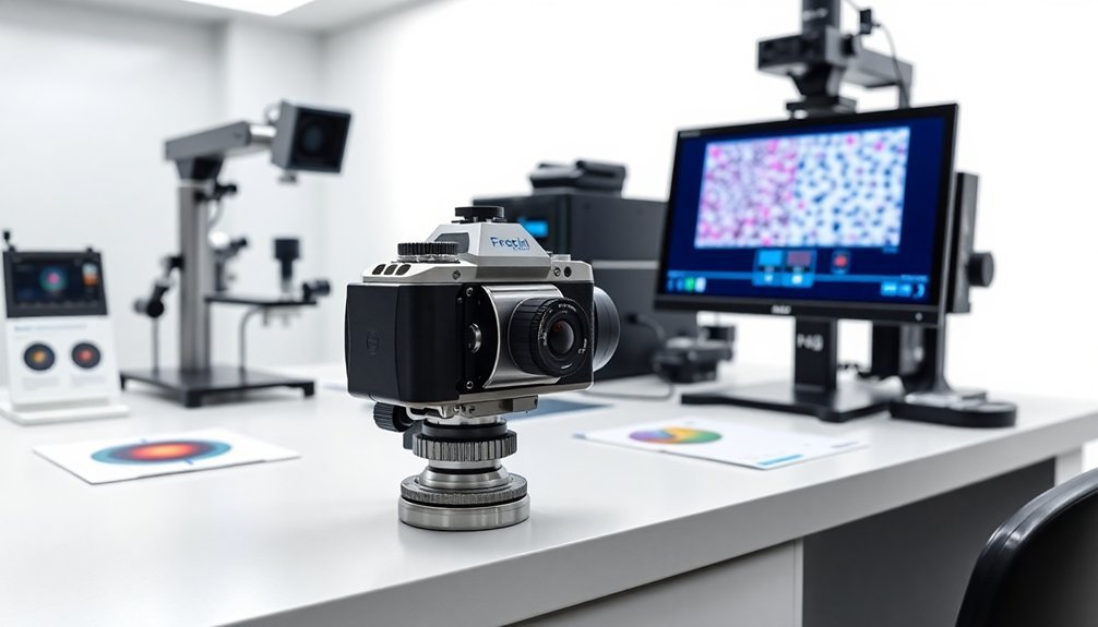
Essential Microscope Camera Interface Protocols
Because microscope camera interfaces form the backbone of digital research imaging, you'll need to master essential protocols like USB3 Vision and Camera Link for ideal data capture. These interoperability standards guarantee your imaging data maintains consistency while enabling seamless communication between devices and software.
| Protocol | Key Benefit |
|---|---|
| USB3 Vision | High-speed transfer |
| Camera Link | Real-time imaging |
| GenICam | Cross-system integration |
| Standard APIs | Software compatibility |
| Custom SDKs | Specialized functions |
When you implement these protocols, you'll enhance your research capabilities through improved collaboration and streamlined workflows. The GenICam standard simplifies your programming tasks across different imaging systems, while maintaining data integrity. This standardization means you can confidently work with equipment from various manufacturers while guaranteeing reproducible results in your scientific studies.
Image Sensor Resolution and Quality Parameters

You'll want to optimize your pixel density by matching the sensor's resolution to your intended output size, with 600 ppi for smaller images and 300 ppi for larger formats.
To maximize dynamic range, capture images in 16-bit mode while maintaining standardized color profiles like AdobeRGB (1998) for consistent reproduction across devices.
When managing noise control, you should implement unsharp masking techniques during post-processing, using radius settings between 0.3 and 0.6 to enhance clarity without compromising image integrity.
Pixel Density Optimization Methods
Since digital imaging continues to advance in scientific research, understanding pixel density optimization has become vital for capturing precise, high-quality data.
To meet current imaging standards, you'll need to balance resolution requirements with practical file management.
Here are key optimization methods to enhance your scientific imaging:
- Adjust your pixel density based on image size: use 600 ppi for 5×7" or smaller, 400 ppi for 8×10", and 300 ppi for images larger than 11×14"
- Implement 16-bit color depth capture to preserve tonal variations and maintain editing flexibility
- Use digital color checker targets for device calibration to guarantee consistent reproduction across different platforms
Remember to maintain efficiency by avoiding unnecessarily large file sizes while making sure you're capturing all essential details for your research requirements.
Dynamic Range Measurement Standards
While dynamic range measurements form the backbone of scientific imaging quality, understanding both sensor resolution and quality parameters is essential for reliable research outcomes.
You'll find that dynamic range standards assess how well your sensor captures both bright and dark areas, directly impacting image quality and detail preservation in your research.
To achieve ideal results, you need to take into account your sensor's resolution, measured by pixel count, alongside vital quality parameters like signal-to-noise ratio for low-light performance.
The modulation transfer function (MTF) helps you evaluate how effectively your sensor reproduces detail and contrast.
When you follow established standards for dynamic range and resolution, you're ensuring consistency in your scientific imaging work, making it easier to interpret and compare research data across different studies.
Noise Control Best Practices
Building on the principles of dynamic range and resolution, effective noise control in scientific imaging requires a thorough approach to sensor technology and quality parameters.
You'll find that higher resolution sensors considerably reduce pixel-level noise artifacts, while proper bit depth selection guarantees better post-processing capabilities.
To maximize your imaging quality and minimize noise, follow these essential steps:
- Select the appropriate sensor technology – CCD sensors typically offer lower noise levels than CMOS sensors for scientific applications.
- Capture images at 16-bit depth to preserve a wider range of tonal values and enhance noise reduction during post-processing.
- Maintain strict environmental controls and regular calibration schedules to optimize sensor performance.
When you're implementing these noise control practices, don't forget to use lossless compression techniques to preserve data integrity while managing file sizes effectively.
Data Management and Storage Specifications

Your data security measures must include robust encryption protocols and role-based access controls to protect sensitive research imaging datasets across all storage environments.
To optimize storage capacity, you'll need to implement tiered storage solutions that balance accessibility with cost-effectiveness, keeping frequently accessed data on high-performance systems while archiving older datasets to more economical platforms.
You should regularly assess your storage requirements based on data growth patterns and retention policies, ensuring compliance with research protocols while maintaining efficient resource utilization.
Data Security Protocols Explained
A robust data security framework serves as the cornerstone of scientific digital imaging protocols.
You'll need to implement thorough measures to protect your research data, particularly when handling sensitive clinical research information.
To maintain data integrity and compliance, follow these essential security protocols:
- Enable encryption for all data, both in transit and at rest, while ensuring your storage solutions provide reliable backup and redundancy features.
- Establish strict access controls through user authentication systems, limiting data accessibility to authorized personnel only.
- Conduct regular security audits to identify potential vulnerabilities and verify compliance with regulations like HIPAA and GDPR.
Storage Capacity Best Practices
Three critical components define effective storage capacity management in scientific digital imaging: scalability, accessibility, and systematic organization.
To maximize your research efficiency, you'll need storage systems that can grow with your data volume, especially when handling multiple clinical trials. Cloud-native solutions offer the flexibility you need, allowing your team to collaborate seamlessly across locations while maintaining robust data security.
Make sure your Research PACS includes advanced metadata management features so you can easily track and retrieve imaging datasets.
You'll want to implement regular audits and access controls to protect data integrity. Don't forget to periodically update your storage infrastructure to accommodate new technologies and prevent data loss.
Calibration Requirements for Scientific Imaging
When conducting scientific research with digital imaging systems, proper calibration stands as the cornerstone of reliable data collection.
You'll need to follow established standards from NARA and NISO while maintaining detailed documentation of your calibration process through machine-readable metadata.
To guarantee your imaging system meets scientific requirements, follow these essential steps:
- Regularly calibrate your cameras using standardized color targets like X-Rite to correct color deviations and maintain consistency across sessions.
- Verify and adjust resolution and bit depth settings to match your specific research needs without creating unnecessarily large files.
- Document each calibration procedure thoroughly, including metadata for each image file, to support transparency and reproducibility.
This systematic approach to calibration will help you produce reliable, high-quality data that meets peer review standards.
Standardized File Formats and Metadata

Building upon proper calibration practices, standardized file formats and extensive metadata form the backbone of scientific imaging protocols.
When you're working with digital imaging systems, you'll find that formats like DICOM enable seamless data exchange between different medical devices and platforms.
You'll need to pay close attention to metadata, as it captures essential information about your imaging process, including device settings and acquisition parameters.
By implementing consistent metadata standards, you can efficiently organize and retrieve your research data, making it easier to manage large datasets for your studies.
Standardized metadata organization empowers researchers to effectively handle and access vast amounts of scientific data for improved study management.
You'll benefit from using machine-readable formats that integrate smoothly with electronic health records and analysis tools.
Remember that adhering to established standards guarantees your images maintain the quality and context necessary for reliable research outcomes.
Quality Control Measures for Image Capture
Since accurate digital imaging is essential for scientific research, implementing robust quality control measures during image capture guarantees the integrity of your data.
In health care and scientific settings, you'll need to follow standardized procedures that guarantee your digitization process remains use-neutral and meets established guidelines.
To maintain consistent quality control throughout your imaging process:
- Verify your file counts and names between directories and spreadsheets, creating high-quality master files that preserve original object characteristics.
- Check metadata components including ICC profiles, resolution, and bit depth to meet required standards.
- Perform visual inspections at 100% magnification to detect artifacts, dirt, or color bias, making minor corrections when needed.
Following FAGDI guidelines, you'll need to implement post-scan adjustments that optimize image quality while maintaining scientific integrity.
Hardware Compatibility Guidelines
You'll need to take into account your legacy system integration requirements when upgrading imaging hardware to guarantee seamless data exchange with existing research infrastructure.
Your hardware must meet current device certification standards, including DICOM compliance and standardized data formats, to maintain compatibility across research environments.
Regular validation of both legacy and new systems against these standards will help you maintain data integrity and research continuity.
Legacy System Integration Requirements
Three critical factors drive the successful integration of legacy systems with modern digital imaging platforms: hardware compatibility, data integrity, and communication protocols.
You'll need to carefully evaluate your existing infrastructure to guarantee seamless operation between old and new components.
To achieve successful integration, focus on these essential requirements:
- Verify your legacy systems' compatibility with DICOM standards and network protocols, implementing necessary adapters or middleware solutions where gaps exist.
- Document all hardware specifications and interface requirements thoroughly, including manufacturer guidelines for both legacy and modern equipment.
- Maintain regular system assessments to identify potential compatibility issues before they impact your research workflow.
You must prioritize data consistency throughout the integration process, guaranteeing your legacy systems can accurately interpret and process information from newer imaging devices.
This approach will help you preserve research integrity while maximizing equipment longevity.
Device Certification Standards
Building upon legacy system requirements, device certification standards establish concrete guidelines for hardware compatibility in digital imaging research. You'll find that these standards guarantee your imaging equipment meets essential performance benchmarks while maintaining seamless integration with HIS and PACS environments.
| Certification Element | Impact on Research |
|---|---|
| DICOM Standards | Guarantees data consistency |
| Hardware Compliance | Enables cross-platform usage |
| Documentation Requirements | Guarantees traceability |
| Performance Validation | Maintains quality control |
To stay current with evolving technology, you must regularly update your certification protocols. When you implement device certification standards correctly, you'll achieve reliable interoperability across your research platforms. Manufacturers must provide detailed documentation proving DICOM standards compliance, while collaborative efforts between regulatory bodies and industry stakeholders continuously refine these requirements to enhance patient safety and data integrity.
Digital Image Processing Standards
While scientific research continues to evolve, standardized digital image processing protocols remain fundamental to guaranteeing data integrity and reproducibility.
You'll find that proper implementation of these standards directly impacts the quality of your research outcomes and the seamless integration of digital imaging across health information systems.
To maintain consistent data quality and interoperability, you should follow these essential processing standards:
- Use lossless compression techniques to preserve original data integrity and guarantee accurate analysis
- Implement thorough metadata management guidelines for efficient organization and retrieval
- Establish continuous feedback mechanisms between clinical users and research teams
When you're working with DICOM standards, you'll need to prioritize consistent data exchange practices.
Color Accuracy and Reproducibility Protocols
Every scientific imaging project demands meticulous color accuracy protocols to secure reproducible results across different systems and laboratories.
You'll need to establish a color-managed environment using device profiles that correct system-specific deviations. Start by implementing digital color checker targets, like X-Rite, to create accurate input profiles for consistent reproduction.
Make certain you're embedding color space information, such as AdobeRGB (1998), in each digital file to maintain color fidelity.
You should regularly verify your color settings and profiles in imaging software to uphold accuracy standards.
Don't forget to document your color models and use color targets consistently – these practices are essential for research reproducibility.
Microscope Camera System Integration
Modern microscope camera systems expand beyond basic color accuracy to provide extensive digital imaging solutions for scientific research.
You'll find these systems seamlessly integrate with your existing imaging software through standardized protocols, making data transfer and analysis more efficient than ever.
To successfully implement DICOM standards in your research workflow, focus on these key aspects:
- Confirm your camera system supports cross-platform compatibility for seamless data sharing across different devices.
- Utilize built-in software features for real-time image processing and live experiment monitoring.
- Take advantage of integrated machine learning capabilities for automated image analysis.
You're now able to conduct more sophisticated research with these advanced systems, as they combine high-resolution imaging with automated analysis tools, streamlining your entire experimental process while maintaining data integrity and accessibility.
Performance Validation Methods
Because scientific research demands unwavering accuracy, validating your digital imaging system's performance is vital for maintaining data integrity. You'll need to conduct regular quantitative assessments using phantom images to evaluate spatial resolution, contrast, and geometric accuracy.
These performance validation methods guarantee your imaging devices meet established benchmarks.
To maintain compliance with Digital Imaging and Communications in Medicine standards, you must implement consistent calibration procedures. You should perform repeatability and reproducibility studies to assess how your imaging systems perform under different conditions.
Don't forget to thoroughly document all validation results and protocol adherence. This documentation isn't just for regulatory compliance—it's essential for verifying the reliability of your research findings and identifying any performance deviations that could compromise your data quality.
Regulatory Compliance in Scientific Imaging
Building upon proper validation protocols, regulatory compliance adds another layer of quality assurance to scientific imaging.
You'll need to understand that DICOM standards are essential for ensuring data exchange and interoperability across imaging systems, though the lack of mandatory compliance remains a significant challenge.
To maintain effective regulatory compliance in your scientific imaging practices:
- Submit manufacturer conformance statements to regulatory bodies like AAO and ARVO, enhancing transparency and standard adherence.
- Stay updated with FDA and National Eye Institute initiatives promoting DICOM standards for patient safety.
- Establish clear timelines for achieving DICOM compliance and track your progress regularly.
You'll find that meeting these requirements not only improves your research quality but also contributes to broader standardization efforts in the scientific imaging community.
Frequently Asked Questions
What Are DICOM Standards?
You'll find DICOM standards are medical imaging protocols that let your healthcare devices communicate and share images effectively. They've been around since 1983, helping guarantee compatibility between different medical imaging systems.
What Is the Principle of Digital Imaging?
You're capturing light that hits electronic sensors, which convert visual information into digital signals. These signals transform into pixels, creating digital images you can process, store, and display on various devices.
When Did DICOM Start?
You'll find that DICOM started in 1983 when the American College of Radiology and National Electrical Manufacturers Association established these standards to create a unified approach for medical imaging communication.
What Is a DICOM License?
You'll need a DICOM license to legally implement DICOM standards in your medical imaging products. It's a mandatory certification that guarantees your devices and software comply with standardized protocols for medical image sharing.
In Summary
You'll find that adhering to digital imaging standards is essential for your research success. By implementing proper camera protocols, maintaining calibration requirements, and following data management specifications, you're ensuring reproducible results. Remember, you're not just capturing images – you're creating reliable scientific evidence. Keep your imaging systems compliant with these standards, and you'll achieve consistent, publication-ready data every time.


Leave a Reply