Modern image sensors are essential for your live specimen analysis because they enable you to capture fast biological processes with exceptional clarity and minimal light exposure. You'll get superior quantum efficiency (up to 95%), ultra-low noise levels (1-3 electrons), and frame rates up to 500 fps, ensuring you won't miss vital cellular events. With features like temperature control and advanced signal processing, you can maintain specimen viability while collecting precise data. Discover how these capabilities transform your research outcomes.
Key Performance Metrics of Modern Image Sensors

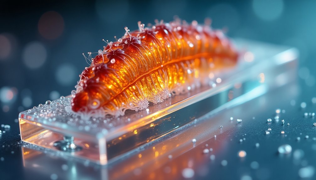
The evolution of modern image sensors has revolutionized live specimen analysis through several vital performance metrics.
You'll find quantum efficiency at the forefront, with back-illuminated devices achieving up to 95% QE for ideal photon collection.
When you're examining sensitivity, read noise becomes essential – sCMOS cameras deliver superior performance with just 1-3 electrons compared to CCD's 5-10 electrons.
You'll need to evaluate pixel size carefully, as larger pixels (like 11 µm) greatly boost your signal-to-noise ratio, though they might affect spatial resolution.
For ideal imaging speed, you'll benefit from modern digital interfaces like PCI Express, which outperforms USB connections in data transfer rates.
These metrics work together to help you capture fast biological processes at up to 500 fps, ensuring you won't miss vital specimen movements.
Real-Time Capture Capabilities for Dynamic Specimens
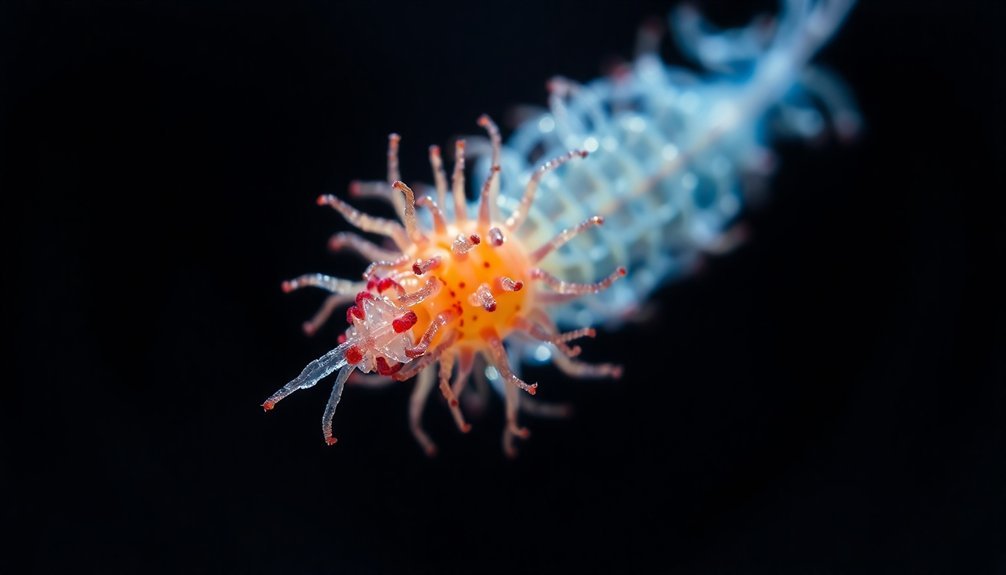
Modern image sensors deliver three essential advantages for capturing dynamic specimens in real time: ultra-fast frame rates up to 500 fps, minimal exposure times, and optimized sensor configurations.
You'll find these capabilities transform live cell imaging through high-speed imaging that reveals previously unobservable biological processes. By reducing exposure time while maintaining a strong signal-to-noise ratio (SNR), you can capture clearer images of dynamic specimens with remarkable precision.
High-speed imaging unveils hidden cellular dynamics while precise exposure control delivers crystal-clear views of specimens in motion.
When you're working with smaller sensor sizes, you'll achieve even faster frame rates – cutting the sensor size in half doubles your imaging speed.
Today's advanced readout systems and PCI Express interfaces boost your data transmission rates, while high sensitivity and low noise features guarantee you'll get sharp, accurate results. These improvements make real-time analysis of dynamic specimens more reliable than ever before.
Resolution and Detail Preservation in Live Imaging
Building on rapid capture capabilities, resolution quality determines your ability to observe minute cellular details in live specimens.
Today's high-resolution image sensors give you unprecedented clarity when examining cellular structures and processes. You'll find that larger pixel sizes greatly enhance detail preservation, especially in low-light conditions where live specimens are most sensitive.
With back-illuminated sensors achieving up to 95% quantum efficiency, you're able to capture even the faintest biological signals with exceptional clarity.
Modern CMOS technology delivers faster readout speeds while reducing noise levels, ensuring you maintain image quality during rapid specimen observation.
When you need extra sensitivity without compromising frame rate, binning techniques let you optimize your imaging setup. These features work together to provide the superior resolution needed for accurate live specimen analysis.
Signal-to-Noise Optimization for Living Samples
You'll need to carefully balance light exposure when imaging live specimens, as excessive illumination can cause photodamage while insufficient light leads to poor signal detection.
To achieve ideal signal-to-noise ratios, you can employ real-time detection strategies that synchronize your camera's exposure timing with your specimen's biological dynamics.
Using high-efficiency sensors and optimized camera settings, you're able to capture clear images with minimal light intensity, protecting your living samples while maintaining strong signal detection.
Minimizing Specimen Light Exposure
When conducting live specimen analysis, minimizing light exposure stands as a critical challenge that demands careful optimization of signal-to-noise ratios.
You'll need to strike a delicate balance between achieving high image quality and protecting your samples from phototoxicity.
To effectively reduce light exposure while maintaining strong SNR, you'll want to leverage cameras with high quantum efficiency (QE), particularly those with back-illuminated sensors.
These devices convert more photons into detectable signals, allowing you to work with lower light intensities.
You can further enhance sensitivity through binning techniques, which combine multiple pixels to boost signal detection.
Additionally, implementing advanced denoising algorithms helps you distinguish true biological signals from background noise, enabling you to work with reduced light levels while preserving essential data quality.
Real-Time Detection Strategies
To achieve reliable real-time detection of biological processes, optimizing signal-to-noise ratio becomes paramount in live specimen analysis.
You'll find that high sensitivity and low noise are essential for capturing accurate biological signals while minimizing unwanted background interference.
You can enhance your signal-to-noise ratio (SNR) by adjusting exposure time and gain settings, while larger sensors improve light collection efficiency by capturing more photons.
When you're dealing with rapid biological changes, modern CMOS sensors offer the acquisition speed you need without compromising image quality.
Advanced denoising algorithms help you combat photon shot noise and camera-related interference, ensuring clearer imaging results.
Temperature Management and Sample Viability
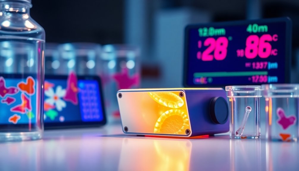
The success of live specimen imaging hinges on precise temperature management. You'll need to maintain your biological specimens at around 37°C to maximize cellular activity and guarantee sample viability. Temperature control systems, like heated stages and incubators, are essential tools you'll want to implement during imaging sessions.
| Temperature Range | Impact | Required Action |
|---|---|---|
| Below 37°C | Reduced cellular activity | Activate heating system |
| At 37°C | Ideal specimen health | Monitor and maintain |
| Above 37°C | Increased phototoxicity risk | Reduce temperature |
Watch out for temperature fluctuations, as they can compromise your experimental results. Remember that high-intensity light sources can elevate temperatures, potentially triggering cellular stress or death. By carefully managing temperature throughout your imaging process, you'll greatly improve the quality and reliability of your specimen analysis.
Frame Rate Impact on Biological Observations
Successful live specimen imaging depends heavily on selecting the right frame rate for your observations. Modern CMOS cameras, capable of reaching 500 fps, let you capture rapid biological events that you couldn't see with older technology.
You'll need to balance frame rate with exposure time; while faster speeds reveal more detail in dynamic processes, they can reduce signal visibility if exposure time is too short.
When you're planning your biological observations, consider how sensor size affects performance. You can achieve higher frame rates by using a smaller imaging area, though you'll sacrifice field of view.
Don't forget about data transmission rates – your camera's interface matters. PCI Express connections will give you faster data transfer than USB, which directly impacts how quickly you can capture and analyze your specimens' behavior.
Quantum Efficiency and Light Sensitivity Considerations
You'll achieve superior results in live specimen imaging by selecting sensors with high quantum efficiency ratings, as these devices can convert up to 95% of incoming photons into measurable electrical signals.
The combination of larger pixel sizes (such as 11 µm) and enhanced QE maximizes your photon capture capability, making it easier to detect faint biological signals from living specimens.
Advanced cooling systems and noise reduction techniques further improve your sensor's light sensitivity, enabling you to capture clear images even under challenging low-light conditions.
Maximizing Photon Capture Rates
When analyzing live specimens, maximizing photon capture rates becomes critical for achieving clear, detailed images while minimizing specimen exposure to light.
You'll want to take into account several key factors to optimize your imaging setup. First, select sensors with high quantum efficiency – back-illuminated sensors can deliver up to 95% QE, greatly improving your signal-to-noise ratio.
To enhance light sensitivity further, you can utilize larger pixel sizes, as an 11 µm pixel captures considerably more light than smaller alternatives. If you're working with particularly challenging specimens, think about implementing 2×2 binning to boost signal capture fourfold.
For dynamic observations, CMOS sensors offer superior readout speeds compared to CCDs, though you'll need to balance exposure time with frame rates to maintain image quality.
Low-Light Detection Thresholds
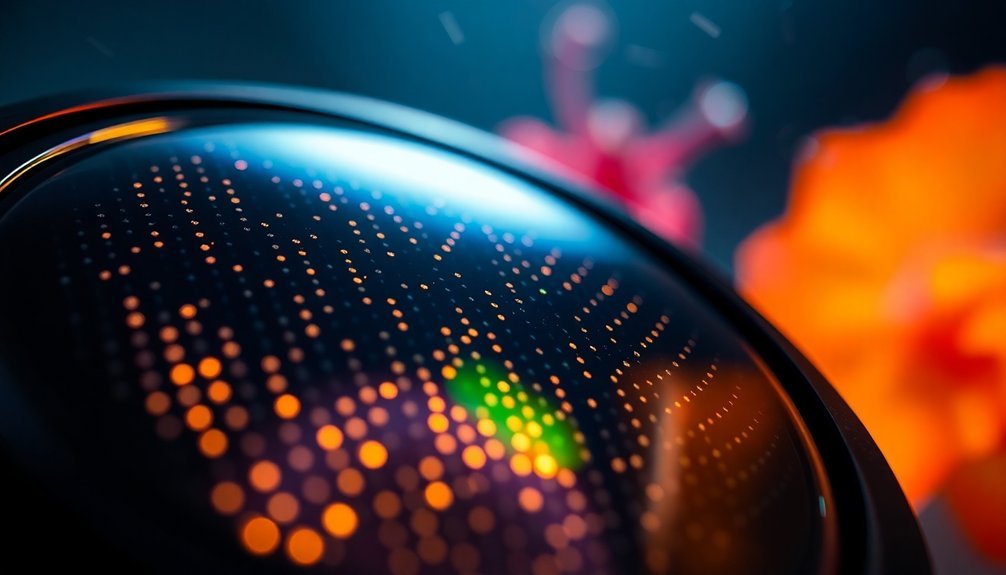
Understanding low-light detection thresholds begins with quantum efficiency and its direct impact on image quality. You'll find that higher quantum efficiency, reaching up to 95% in modern sensors, directly improves your low-light detection capabilities during imaging experiments.
- Signal-to-noise ratio (SNR) determines how clearly you'll detect faint signals against background noise, vital for live specimen analysis.
- Larger pixels (11 µm vs 4.5 µm) capture more light, boosting your sensor's high sensitivity in challenging conditions.
- Read noise and dark current noise greatly affect detection limits, with top cameras achieving just 1-3 electrons of read noise.
- Cooling systems reduce dark current, helping you achieve better results in low-light scenarios.
When you're working with dynamic biological processes, these factors combine to determine your practical detection threshold and overall imaging success.
Sensor Architecture Effects on Sample Analysis
The architecture of an image sensor plays a vital role in determining the quality and accuracy of live specimen analysis.
You'll find that CMOS sensors offer faster readout times, enabling you to capture rapid biological processes, while back-illuminated sensors maximize quantum efficiency up to 95%, helping you detect faint fluorescence signals.
When you're working with low-light applications in live cell imaging, the sensor architecture's pixel design becomes essential – larger pixels enhance sensitivity, and binning capabilities let you combine multiple pixels for improved light detection.
For real-time analysis of dynamic specimens, you'll benefit from high-speed digital interfaces like PCI Express, which guarantees rapid data transfer from sensor to processor.
These architectural features work together to provide you with the precision and speed needed for thorough specimen observation.
Field of View and Sample Coverage Trade-offs
Successful live specimen analysis hinges on balancing field of view (FOV) and sample coverage requirements.
You'll need to evaluate how sensor design impacts your imaging applications, as the trade-off between FOV and spatial resolution directly affects your results.
- Your specimen's size and movement patterns determine the required field of view, influencing your choice of sensor dimensions.
- Pixel size impacts your ability to capture fine details, with larger pixels typically offering better sensitivity but reduced resolution.
- You'll need to assess whether sample coverage or detail is more critical for your specific application.
- The trade-off between FOV and resolution can be partially mitigated by selecting sensors with suitable pixel density.
When choosing your imaging setup, remember that maximizing FOV often means compromising resolution unless you're using larger sensors that maintain both coverage and detail.
Digital Processing and Data Management Strategies
While capturing high-quality images of live specimens is essential, effective digital processing and data management play equally important roles in achieving meaningful results.
You'll find that digital signal processing enhances high sensitivity and low noise characteristics, improving your signal-to-noise ratio (SNR) through real-time adjustments during specimen analysis.
To handle the massive volumes of high-resolution imaging data you'll generate, you'll need robust data management strategies.
Consider implementing cloud-based data management platforms that enable seamless collaboration with colleagues worldwide. You can optimize your workflow by using machine learning algorithms to detect specific biological features and monitor dynamic changes.
Don't forget to employ image compression techniques – they'll help you manage storage space efficiently without compromising the quality of your specimen imaging data.
Frequently Asked Questions
Why Are Image Sensors Important?
You'll find image sensors essential as they convert light into digital signals, helping you capture high-quality images with excellent sensitivity, reduced noise, and fast frame rates for analyzing dynamic processes and microscopic details.
What Is the Function of the Image Sensor?
You'll find that image sensors work by converting light from your specimen into electrical signals that create digital images. They'll capture and transform these photons, letting you analyze biological processes in real-time.
How Does a Sensor Affect Image Quality?
Your sensor's sensitivity, quantum efficiency, and noise levels directly impact how clearly you'll see details. Better sensors reduce background noise, capture more light, and deliver sharper images with higher signal-to-noise ratios.
Why Is Sensor Sensitivity Important?
You'll need high sensor sensitivity to detect faint signals from live specimens, ensuring you don't miss critical details. It's essential for capturing clear images in low-light conditions while minimizing sample damage.
In Summary
You'll find that choosing the right image sensor is essential for successful live specimen analysis. By carefully considering factors like quantum efficiency, thermal management, and real-time capture capabilities, you're ensuring ideal results while maintaining sample viability. Remember, it's not just about resolution – you need to balance sensitivity, speed, and field of view to capture the dynamic nature of living specimens effectively.


Leave a Reply