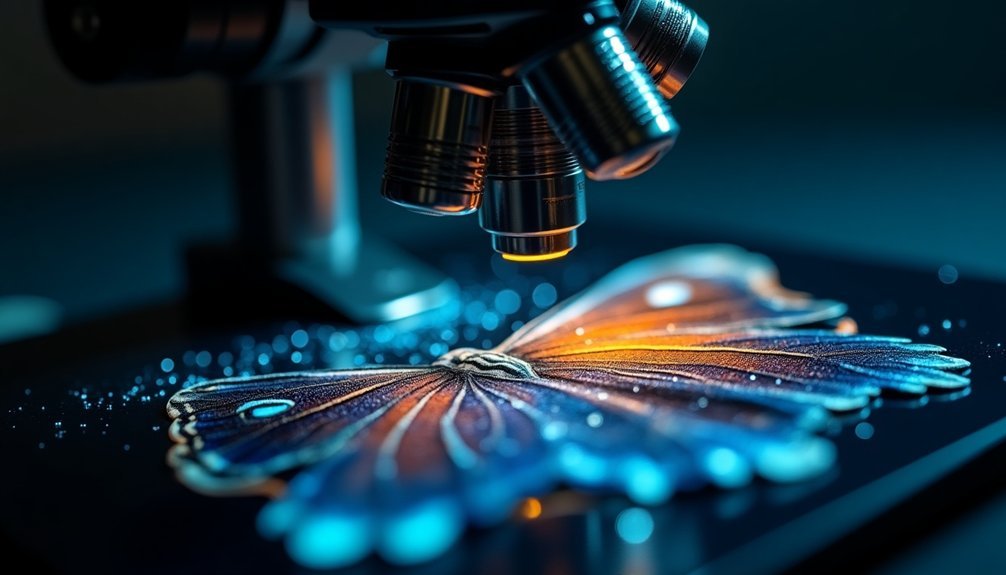
You'll find several powerful software options to enhance your digital microscope images. ImageJ and Adobe Photoshop offer basic sharpening and contrast improvements, while specialized tools like Huygens and AutoQuant provide advanced deconvolution capabilities. AI-powered enhancement software can transform low-quality captures into crystal-clear visualizations through deep learning algorithms. For real-time improvements, modern microscope systems integrate with software that provides instant optimization. Understanding these tools will reveal new levels of microscopic detail and clarity.
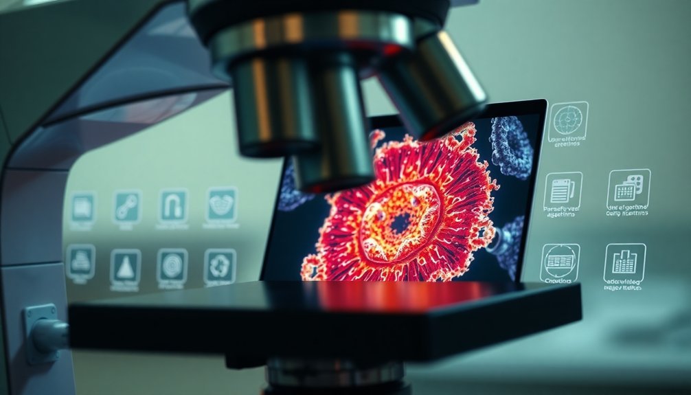
Resolution Enhancement Algorithms and Technologies
Several powerful resolution enhancement techniques transform low-quality microscope images into sharper, more detailed outputs.
When you're working with a digital microscope, you'll find that resolution enhancement algorithms can greatly boost your image quality by increasing pixel count and reconstructing fine details.
You can leverage deep learning methods like Point-Scanning Super-Resolution (PSSR), which uses trained models to convert low-resolution inputs into high-resolution images.
Image processing tools also offer image stacking capabilities, combining multiple focal plane shots for enhanced depth and clarity.
Advanced software features in programs like Adobe Photoshop and ImageJ help you sharpen images and improve contrast.
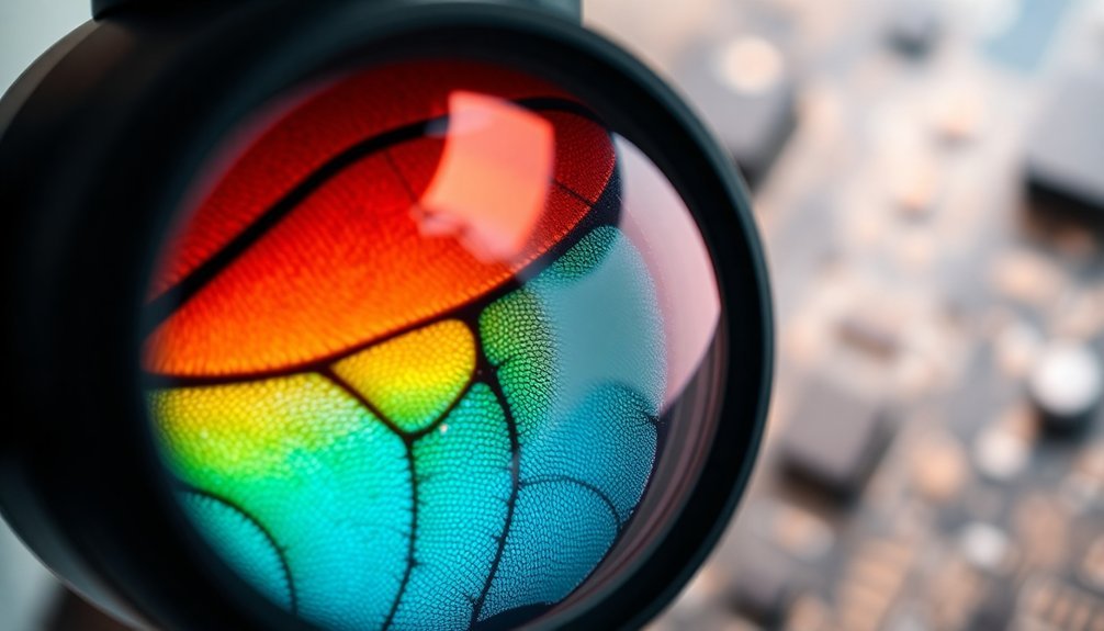
Additionally, edge detection algorithms emphasize structural details in your microscopic samples, making critical features more visible and easier to analyze.
Image Processing and Deconvolution Solutions
Modern deconvolution software tools like Huygens and AutoQuant will sharpen your microscope images by reversing optical blurring effects and reducing unwanted noise.
You'll find these applications particularly effective at reconstructing high-resolution details from multiple low-resolution frames through super-resolution techniques.
Your image processing workflow can be streamlined through batch processing features, allowing you to enhance large sets of microscopy data while maintaining consistent quality across all samples.
Advanced Deconvolution Software Tools
Advanced deconvolution software tools have revolutionized digital microscopy by transforming blurry, low-quality images into sharp, detailed representations. When you're working with high-resolution microscopic imaging, you'll find that these tools can improve the quality of your results by up to 2-3 times.
Software like Huygens and AutoDeblur uses sophisticated algorithms to model your imaging system's point spread function (PSF), helping you achieve clearer, more accurate image analysis.
Key benefits you'll experience with advanced deconvolution software:
- Enhanced visibility of fine structural details in biological samples, critical for histology and cellular research
- User-friendly interfaces with customizable settings to optimize your specific microscopy applications
- Powerful reconstruction capabilities that reverse optical imperfections and reduce noise in your images
Real-Time Enhancement Algorithms
While traditional microscopy relied on post-processing techniques, real-time enhancement algorithms now transform your microscope images instantly as you work. These powerful tools dynamically adjust contrast and sharpness during your live imaging sessions, giving you clearer, more detailed views without delay.
Modern digital microscopy software packages integrate sophisticated algorithms that tackle optical imperfections head-on. You'll find that these solutions use mathematical models to convert lower-resolution data into high-resolution images, all without requiring extra hardware.
The real-time enhancement algorithms work alongside deconvolution processes to reverse blurring effects and boost overall image quality.
You'll save significant time by reducing post-processing needs, as these tools let you analyze and interpret your microscopic images faster than ever before. Your research workflow becomes more efficient with instant visual improvements.
Machine Learning and AI-Powered Enhancement Tools
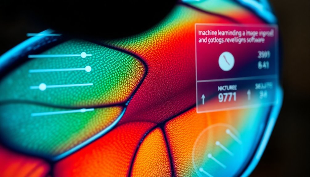
Modern AI-powered microscopy tools now offer you remarkable image enhancement capabilities through deep learning algorithms that can transform low-quality images into crisp, detailed visualizations.
You'll find real-time resolution optimization features that instantly process and improve your microscope feed, making cellular structures more visible even under challenging conditions.
These systems require extensive training on paired low and high-resolution image datasets, but once implemented, they'll considerably reduce your need for expensive hardware while maintaining professional-grade image quality.
Deep Learning Enhancement Features
Recent breakthroughs in deep learning have revolutionized digital microscope image enhancement, particularly through convolutional neural networks (CNNs). You'll find that modern imaging software can transform low-quality microscopy images into high-resolution outputs by learning from extensive training datasets.
The innovative "Crappifier" tool and PSSR software make this technology more accessible, helping you achieve better results without expensive equipment.
- Deep learning algorithms analyze patterns and details from high-quality reference images to enhance poor-quality captures
- Advanced imaging software reduces your dependence on costly high-resolution microscopes while maintaining excellent output quality
- The combination of AI and microscopy speeds up your workflow while preserving essential scientific details
These features aren't just improving image quality – they're making advanced microscopy techniques more accessible and efficient for researchers across scientific disciplines.
Real-Time Resolution Optimization
As microscopy technology advances, real-time resolution optimization has become a game-changing feature in digital imaging software.
You'll find that these tools use deep learning techniques to enhance image quality during live sessions, eliminating the need for extensive post-processing work.
When you're working with digital microscopes, real-time resolution optimization tools can help you observe cellular structures under low-light conditions with remarkable clarity.
They'll analyze your low-resolution images and intelligently reconstruct missing details, producing high-resolution results instantly.
Solutions like Point-Scanning Super-Resolution (PSSR) effectively improve naturally poor-quality images by learning from both high-resolution and artificially degraded samples.
This technology means you won't need expensive equipment to achieve quality results, making advanced microscopy more accessible and efficient for your research needs.
AI Training Requirements
Training AI-powered enhancement tools for digital microscopy requires extensive high-resolution image datasets to achieve ideal results.
You'll find that software offers innovative solutions to overcome data limitations, like the "crappifier" tool that artificially degrades high-quality images to create training pairs. This approach helps you develop robust deep learning models without needing massive collections of natural low-quality images.
- Point-Scanning Super-Resolution (PSSR) models learn by comparing high-resolution and degraded digital image pairs
- The Salk Institute's method makes AI training more accessible by artificially creating low-quality versions of existing images
- Deep learning solutions reduce your dependence on expensive high-resolution microscopy equipment
These AI training requirements, while demanding, ultimately lead to more cost-effective and accessible microscopy tools for your research needs.
Real-Time Image Optimization Software
While digital microscopy continues to evolve, real-time image optimization software has become essential for achieving superior resolution and clarity during live specimen observation.
You'll find this technology seamlessly integrates with your microscope system, providing instant feedback and adjustments that eliminate lengthy post-processing work.
With an intuitive user interface, you can capture high-resolution images using auto-exposure features and manual fine-tuning options, regardless of lighting conditions.
The software's advanced capabilities include live image stacking, which combines multiple focal plane images to create a single, detailed view with enhanced depth of field.
Whether you're new to digital microscopy or an experienced user, you'll appreciate how real-time image optimization software streamlines your workflow while delivering superior image quality and resolution.
Advanced Stacking and Reconstruction Methods
Building upon real-time optimization capabilities, advanced stacking and reconstruction methods take digital microscopy resolution to new heights.
You'll find that advanced stacking techniques, like those in EoDF, combine multiple focal depth images to create sharper, more detailed results. Image stacking methods align and merge several captures, effectively reducing noise while enhancing clarity.
With software tools like Adobe Photoshop, ImageJ, and VIAS, you can transform your microscopy data into high-resolution 3D visualizations.
- Use image reconstruction to reveal intricate structural details that aren't visible in single captures
- Combine multiple focal planes to achieve superior depth perception and overall image quality
- Leverage specialized software to align and integrate complex microscopy stacks into coherent datasets
Hardware-Software Integration for Quality Improvement
Since digital microscopy relies on seamless hardware-software integration, you'll need to understand how these components work together to maximize image quality. Your software controls Microscope Cameras and enhances Resolution through specialized tools like LAS X, which lets you adjust exposure settings for ideal clarity. Digital microscopes like the PreciPoint M8 combine advanced hardware with sophisticated software to deliver superior image quality.
| Component | Hardware Role | Software Integration |
|---|---|---|
| Camera | Image Capture | Exposure Control |
| Lighting | Illumination | Auto-adjustment |
| Focus | Focal Planes | Image Stacking |
| Resolution | Sensor Quality | Digital Enhancement |
| Analysis | Data Collection | ImageJ/Cell Profiler |
You'll achieve the best results when your software precisely controls microscope settings while utilizing features like image stacking and quantitative analysis tools to process and enhance your captured images.
Frequently Asked Questions
How to Improve Microscope Image Quality?
You'll improve microscope image quality by optimizing lighting angles, using image stacking techniques, adjusting focus depth, and applying post-processing tools to enhance clarity, contrast, and sharpness in your captured images.
How to Increase Resolution on a Microscope?
You'll achieve higher microscope resolution by using high NA objectives, optimizing lighting conditions, and utilizing image stacking features. Don't forget to properly adjust DPI settings and employ quality digital microscopy software.
What Improves Resolution of an Image Seen Under a Microscope?
You'll get better microscope resolution by using higher numerical aperture objectives, shorter wavelength light, and proper focus adjustment. Image stacking and deconvolution software can also help enhance the clarity of your microscopic views.
How Do You Make a Microscope Image Clearer?
You'll get clearer microscope images by optimizing lighting angles, using image stacking software, maintaining clean lenses, and ensuring proper calibration. Don't forget to use high-resolution settings during capture for better results.
In Summary
You'll find multiple software solutions to enhance your digital microscope images. From deconvolution algorithms and AI-powered tools to real-time optimization software, these technologies can greatly improve resolution quality. Consider integrating stacking methods and machine learning applications with your existing hardware setup. When properly implemented, these tools will transform your low-resolution microscope images into clearer, more detailed representations of your specimens.


Leave a Reply