Essential stage accessories for time-lapse cell imaging include environmental control systems maintaining 37°C temperature and appropriate gas levels, precision motorized stages with anti-drift technology, specialized vessel holders for different labware, and anti-vibration solutions. You’ll need focus maintenance systems like hardware autofocus to prevent blur during extended sessions. Software integration enables automated coordination between components while minimizing phototoxicity through illumination controls. These accessories work together to produce consistent, high-quality cellular imaging data throughout your experiments.
Environmental Control Systems for Live Cell Preservation
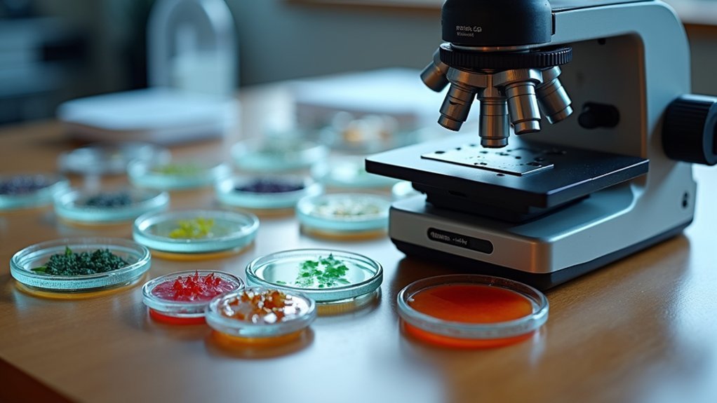
When conducting time-lapse imaging of living cells, maintaining physiological conditions is absolutely critical for experimental success.
Environmental control systems like the EVOS Onstage Incubator regulate temperature, humidity, and gas levels to create ideal conditions for your samples during extended observation periods.
Maintain optimal cellular microenvironments for extended imaging with precise temperature, humidity and gas regulation.
You’ll benefit from the EVOS system’s precise temperature control, maintaining 37°C with 70-90% humidity—essential parameters for cell viability.
The compact environmental chamber (27 x 17 x 4.1 cm) accommodates various vessels while saving valuable lab space.
What makes these systems indispensable for live-cell imaging is their ability to adjust parameters mid-experiment without disrupting your samples.
This flexibility, combined with contamination prevention features, guarantees reliable time-lapse imaging results while maintaining cell health throughout your observation window.
Precision Motorized Stages for Sequential Imaging
Precision motorized stages enhance your time-lapse imaging with multi-point position control, allowing you to capture sequential data from various samples simultaneously.
You’ll benefit from integrated temperature stability features that prevent thermal drift and maintain consistent cell conditions throughout extended imaging periods.
Modern systems offer seamless software integration capabilities, enabling you to program complex acquisition sequences and synchronize stage movements with your imaging parameters.
Multi-Point Position Control
To effectively capture cellular dynamics across multiple regions of interest, precision motorized stages have become indispensable components in time-lapse imaging setups.
These sophisticated platforms allow you to program multiple positions with exceptional accuracy, ensuring you’ll return to the exact same cells throughout your experiment.
With multi-point position control, you’re able to monitor numerous samples simultaneously without manual intervention.
Your motorized stages work seamlessly with autofocus systems, maintaining ideal focus at each predetermined location while compensating for any drift that might occur during extended time-lapse cell imaging sessions.
You’ll appreciate the significant efficiency gains as automated movement between positions eliminates tedious manual repositioning.
This technology is compatible with various labware types on inverted microscopes, giving you versatility while ensuring consistent imaging conditions across all your experimental sites.
Temperature Stability Features
Maintaining consistent temperature throughout your time-lapse experiments is critical for capturing authentic cellular behaviors. Precision motorized stages contribute greatly to temperature stability by minimizing vibrations and eliminating the need for manual adjustments that could disrupt your carefully controlled environment.
| Feature | Benefit | Application |
|---|---|---|
| Micrometer-level positioning | Eliminates drift | Long-duration studies |
| Autofocus integration | Reduces manual intervention | Temperature-sensitive samples |
| Vibration dampening | Maintains stable environment | High-magnification imaging |
| Incubator compatibility | Guarantees consistent conditions | Live cell experiments |
When selecting imaging systems for time-lapse studies, prioritize motorized stages that offer precise control over sample positioning. These stages work seamlessly with environmental chambers to maintain temperature stability throughout your experiment, preventing thermal fluctuations that could compromise your results or introduce artifacts into your cellular behavior data.
Software Integration Capabilities
Seamless software integration represents the cornerstone of effective time-lapse imaging workflows when using precision motorized stages.
You’ll find that modern motorized stages can be synchronized with diverse software solutions, allowing for automated coordination between stage movements and image acquisition during your experiment setup.
When you implement integrated imaging software with your motorized stages, you’ll benefit from programmable controls that let you precisely define acquisition parameters, including capture intervals and movement paths across your samples.
This integration minimizes human error while maximizing reproducibility in your time-lapse studies.
For best results, choose systems with autofocus compatibility, as this guarantees consistent image quality throughout extended acquisition periods.
The right software-stage combination will transform your imaging workflow, providing reliable data collection with minimal manual intervention.
Vessel Holders and Stage Plates Optimized for Cell Culture
You’ll find both universal vessel holders that accommodate multiple formats and custom holders designed for specific culture vessels, each offering distinct advantages for your imaging workflow.
Temperature stability features in premium stage plates prevent thermal gradients across your samples, ensuring consistent cellular responses during extended time-lapse experiments.
When selecting holders for multi-well plates, consider compatibility with your microscope’s automated positioning system to maximize efficiency in sequential imaging of multiple samples.
Universal vs. Custom Holders
When designing your time-lapse imaging setup, choosing between universal and custom holders represents a critical decision that impacts experimental flexibility and precision.
Universal holders offer versatility, accommodating various labware types from microscope slides to culture flasks, giving you adaptability across different experimental protocols.
However, custom holders provide optimized support for specific brands and vessel types, ensuring precise alignment during imaging that can’t be matched by universal options.
For live-cell imaging, EVOS vessel holders are specifically engineered to minimize optical issues like autofluorescence that might compromise your results.
If you’re using the EVOS Onstage Incubator, you’ll need compatible holders to maintain proper environmental control.
For time-lapse experiments on the EVOS M5000’s Stage View feature, custom holders deliver the stability necessary for consistent, high-quality imaging throughout your experiment.
Temperature Stability Features
Maintaining consistent temperature throughout your time-lapse imaging experiments represents one of the most critical requirements for accurate cell behavior monitoring.
EVOS vessel holders are specifically engineered with temperature stability features that minimize fluctuations during extended live-cell imaging sessions.
When you select compatible holders for your EVOS Onstage Incubator, you’ll guarantee peak thermal conductivity while preventing optical issues that could compromise your results.
These specialized accessories accommodate various brands of plates, flasks, and dishes while facilitating precise monitoring of experimental conditions.
The Stage View feature of the EVOS M5000 works seamlessly with these holders to track cellular changes over time.
The right vessel holder not only enhances alignment precision but also reduces contamination risks and maintains the humidity conditions essential for keeping your cultures viable during long-term observations.
Multi-well Compatibility Options
EVOS imaging solutions accommodate a vast array of multi-well vessels, guaranteeing your time-lapse experiments proceed without interruption.
The specialized EVOS vessel holders and stage plates are engineered to fit multiple brands of plates, flasks, and dishes with precision alignment that enhances your imaging workflow.
These accessories greatly improve your results by reducing optical issues like autofluorescence that often plague standard plastic labware.
When conducting live-cell imaging with the EVOS Onstage Incubator, you’ll appreciate the versatility to use T-25 flasks, chamber slides, and various multi-well plates without compromising ideal incubator operation.
For consistent time-lapse results, always select the appropriate vessel holder from the available options.
This tailored approach guarantees your specific experimental requirements are met while maintaining imaging accuracy throughout extended observation periods.
Integrated Gas Regulation Systems for Cellular Homeostasis
The success of time-lapse cell imaging experiments often depends on precise control of atmospheric conditions that mirror physiological environments. Integrated gas regulation systems in the EVOS Onstage Incubator provide this essential capability, maintaining exact levels of O₂, CO₂, and nitrogen to support cellular homeostasis during live-cell imaging experiments.
You’ll find the OSI-2 model particularly valuable for its user-defined controls that enable accurate gas concentration adjustments. These systems create specific environmental conditions ideal for hypoxia studies and other gas-sensitive cellular processes while minimizing cellular stress during extended imaging sessions.
The ability to monitor and adjust gas levels in real-time streamlines your experimental workflow, freeing you to focus on data collection rather than environmental maintenance. This precision ultimately enhances the physiological relevance and reliability of your research results.
Anti-Vibration Solutions for Image Stability
When capturing subtle cellular dynamics over extended periods, vibration can become your experiment’s worst enemy.
Even minor tremors can blur images and compromise your time-lapse cell imaging results, making anti-vibration solutions essential investments.
Anti-vibration tables provide vital isolation from environmental disturbances through their heavy, dampened structures.
You’ll notice a significant improvement in your signal-to-noise ratio, resulting in clearer, more precise imaging data.
Most tables feature adjustable feet to compensate for uneven surfaces, ensuring stability regardless of your lab setup.
Beyond improving image quality, these solutions protect your equipment by reducing mechanical stress on sensitive components.
Focus Maintenance Technologies for Extended Imaging Sessions
Maintaining precise focus throughout extended time-lapse imaging presents considerable challenges as thermal drift, stage settling, and environmental fluctuations can gradually shift your focal plane.
To overcome these obstacles, you’ll need reliable focus maintenance technologies that actively compensate for these changes.
Motorized focus systems automatically adjust focus during long sessions, counteracting temperature-induced position shifts.
Both hardware and software-based autofocus systems continuously monitor and correct focus, ensuring ideal clarity throughout your experiment.
When paired with sensitive cameras, these technologies help maintain high signal-to-noise ratios critical for detailed cellular imaging.
For best results, install your focus maintenance equipment on anti-vibration tables to minimize external disturbances.
Remember to regularly calibrate your systems—even minor focus shifts can greatly impact time-lapse imaging quality during experiments spanning hours or days.
Illumination Controls for Minimizing Phototoxicity
Effective illumination control stands at the center of successful time-lapse cell imaging, as excessive light exposure can trigger phototoxic damage that compromises your experiment’s validity.
Light management is the cornerstone of time-lapse imaging—prevent phototoxicity or risk experimental failure.
To mitigate these risks, incorporate specialized shutters and adjustable field diaphragms that reduce cells’ exposure time to intense light.
Pair your setup with sensitive cameras featuring high signal-to-noise ratios, allowing you to decrease excitation light intensity while maintaining image quality.
You’ll also want to optimize recovery periods between exposures, giving cells time to repair between imaging sequences.
Consider adding antioxidants to your culture medium to combat oxidative stress during live-cell imaging.
Motorized stages with automated focus capabilities enable sequential imaging at multiple positions, minimizing overall light exposure while maximizing observation opportunities.
These strategies collectively preserve cellular integrity throughout your extended imaging sessions.
Digital Integration Platforms for Experiment Monitoring
Modern digital integration platforms serve as the command center for sophisticated time-lapse imaging experiments, eliminating the need for constant manual supervision.
Systems like the EVOS Onstage Incubator maintain precise physiological conditions by regulating temperature, humidity, and gas levels—critical factors for accurate live-cell imaging results.
You’ll appreciate the intuitive interfaces that simplify experiment setup and monitoring, allowing real-time parameter adjustments without disturbing ongoing observations.
When integrated with imaging systems such as the EVOS M5000 or M7000, these platforms enable seamless data capture over extended periods.
The compatibility with specialized vessel holders and stage plates further enhances imaging quality by minimizing optical issues.
This thorough approach to environmental control guarantees your time-lapse studies capture authentic cellular processes under ideal conditions.
Frequently Asked Questions
How Often Should Stage Accessories Be Calibrated for Optimal Performance?
You should calibrate stage accessories every 3-6 months or before critical experiments. Regular calibration guarantees accuracy, prevents drift, and maintains precision. Don’t wait until problems arise—proactive maintenance yields reliable results.
Can Consumer-Grade Cameras Be Adapted for Time-Lapse Cell Imaging?
You can adapt consumer cameras for time-lapse cell imaging with proper modifications. You’ll need microscope adapters, remote triggers, and software for interval shooting. However, they won’t match specialized scientific cameras’ sensitivity.
What Emergency Backup Systems Prevent Data Loss During Power Outages?
You’ll need UPS systems, automatic data backup to cloud servers, redundant storage, and generators to prevent data loss during outages. They’ll keep your imaging systems running and safeguard your time-lapse sequences automatically.
How Do Different Cell Lines Affect Accessory Selection Criteria?
Different cell lines require specific accessories based on their growth characteristics. You’ll need custom chambers for suspension cells, specialized surfaces for adherent cells, and temperature controls matching each line’s ideal growth conditions.
Are There Portable Stage Systems Suitable for Educational Demonstrations?
Yes, you’ll find several portable stage systems ideal for classroom demonstrations. They’re compact, lightweight, and offer basic temperature control with simplified interfaces that students can easily understand during educational cell biology presentations.
In Summary
Your success with time-lapse cell imaging hinges on these essential accessories. You’ll need proper environmental control, precision stages, and specialized holders to maintain cellular health. Don’t overlook vibration dampening and focus maintenance technologies that guarantee image quality. By incorporating gas regulation systems and controlling illumination to reduce phototoxicity, you’re well-equipped to capture cellular dynamics with unprecedented clarity and consistency.


Leave a Reply