Time-lapse videos of dividing cells showcase life’s microscopic miracles, condensing hours into seconds. You’ll be amazed by frog embryo development transforming from four cells to millions in just 23 seconds. Modern microscopy techniques with specialized LED lighting and anti-vibration tables capture every detail of mitosis. These videos highlight prophase, metaphase, anaphase, and telophase with stunning clarity. This fusion of art and science reveals nature’s hidden choreography in mesmerizing detail.
5 Best Time-Lapse Videos of Dividing Cells
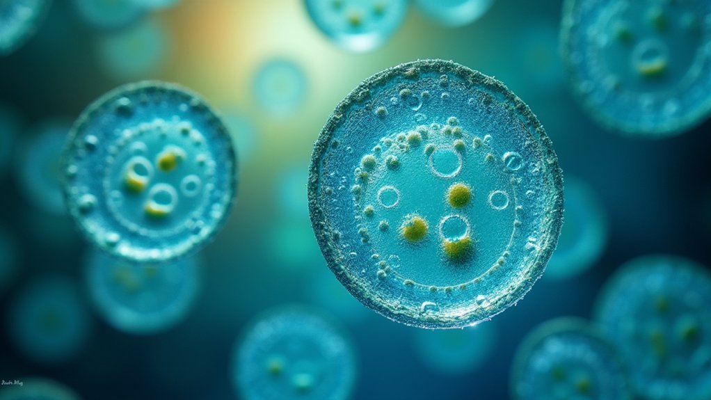
Have you ever wondered what the miracle of life appears like at the cellular level? Search no more than the remarkable time-lapse video of cell division in the common frog, Rana temporaria. This stunning footage condenses 33 hours of development into just 23 seconds, showing the transformation from four cells to millions.
What makes these videos so extraordinary is the specialized microscopy used to capture them. Scientists utilize customized microscopes with infinity optical systems, precise lighting, and an anti-vibration table to guarantee crystal-clear imagery. The result resembles computer-generated effects, but it’s entirely natural.
These time-lapses aren’t just visually impressive—they’re valuable educational tools that reveal the dynamic complexity of embryonic development, inspiring greater interest in developmental biology and microscopy techniques.
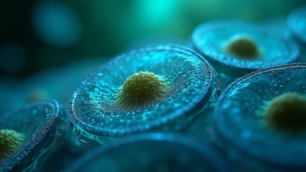
Frog Embryo Development: From Zygote to Tadpole
While most people only see tadpoles swimming in ponds, the journey from a single cell to a complex organism is nothing short of extraordinary.
Time-lapse videos reveal how a single zygote of the common frog transforms through rapid cell division over just 33 hours.
You’ll witness the fascinating progression from cleavage to blastula formation, followed by gastrulation and neurulation. As organogenesis begins, the heart and nervous system take shape within the transparent eggs.
The videos capture cellular differentiation that’s invisible to the naked eye, highlighting how a simple cell evolves into a complex tadpole.
This remarkable transformation, occurring in thousands of eggs laid in shallow freshwater, offers invaluable insights into early amphibian development that would otherwise remain hidden from view.
Revolutionary Microscopy Techniques Behind Cell Division Footage
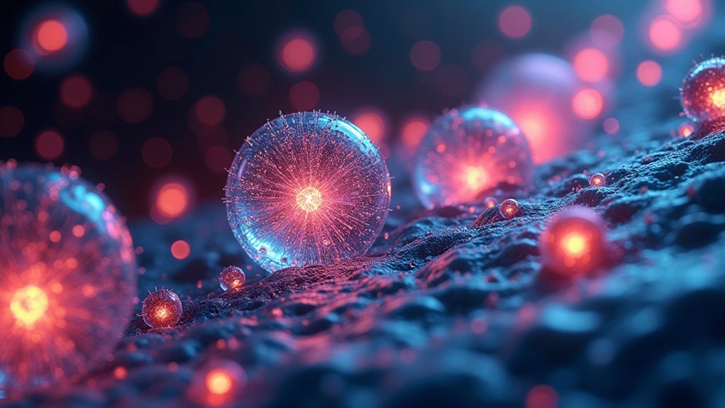
The breathtaking footage of frog embryo development wouldn’t be possible without cutting-edge microscopy innovations that transform scientific observation into visual art.
These remarkable videos employ customized infinity optical designs and specialized LED lighting to capture cellular details so precise they resemble CGI.
To achieve this clarity, scientists place microscopes on anti-vibration tables that eliminate disruptive movements while precisely controlling ambient light and temperature. This meticulous setup allows them to document rapid cellular changes with extraordinary stability.
Perhaps most impressive is how these advanced imaging techniques compress time itself—condensing a 33-hour division process into just 23 seconds of enchanting footage.
You’re not just watching educational content; you’re experiencing biological artistry made possible through revolutionary technology that reveals nature’s hidden choreography in unprecedented detail.
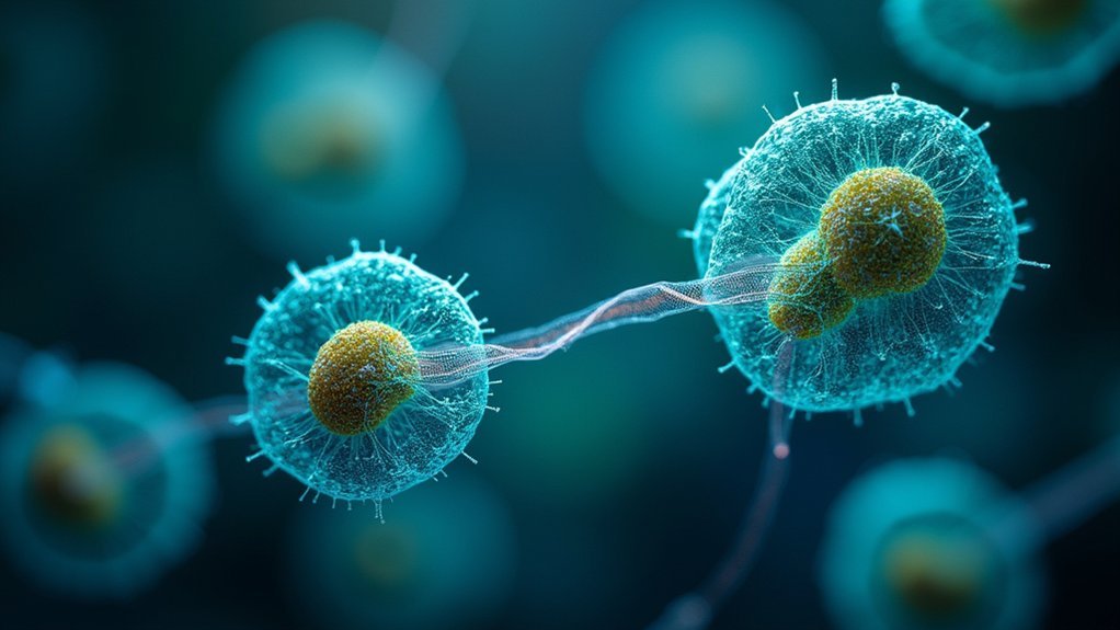
Capturing the Dance of Chromosomes: Mitosis in High Definition
Mesmerizing in their choreographed precision, high-definition time-lapse videos of mitosis reveal nature’s most spectacular cellular performance. You’ll witness hours of complex cellular activity condensed into seconds, making the intricate stages of cell division more accessible and understandable.
| Mitosis Stage | What You’ll Observe |
|---|---|
| Prophase | Chromosomes condensing, nuclear envelope breaking down |
| Metaphase | Chromatid pairs aligning at the cell’s equator |
| Anaphase | Sister chromatids separating and moving to opposite poles |
| Telophase | Nuclear envelopes reforming around new chromosome sets |
Fluorescent tagging techniques enhance these videos, highlighting chromosomes against cellular backgrounds. You’ll appreciate not just the biology but also the artistry of cell division—a perfect educational tool that transforms complex cellular processes into visual poetry.
Cellular Symphony: The Art and Science of Time-Lapse Biology
Beyond the detailed observation of mitosis lies a broader artistic dimension where cellular biology transforms into visual symphonies.
You’re witnessing nature’s complexity condensed into stunning moments when you watch a frog egg’s 33-hour division journey compressed into just 23 seconds.
The marriage of technology and biology creates these visual feasts. Custom lenses and LED lighting enhance microscopic details that would otherwise remain hidden, revealing cellular changes with breathtaking clarity.
These videos aren’t merely scientific documentation—they’re powerful educational tools that engage your curiosity.
What makes time-lapse biology so compelling is this perfect fusion of art and science. The visual impact of rapidly dividing cells inspires viewers to explore developmental biology further while showcasing the intricate beauty underlying life’s fundamental processes.
Frequently Asked Questions
What Are the Most Rapidly Dividing Cells?
You’ll find bone marrow’s hematopoietic stem cells divide every 24 hours. Bacteria divide even faster—E. coli can reproduce every 20 minutes! Skin cells turnover monthly, while some cancer cells divide uncontrollably at alarming rates.
What Is the Fastest Dividing Cell?
E. coli bacteria are the fastest dividing cells, replicating every 20 minutes under ideal conditions. You’ll find this rate considerably outpaces even your rapidly dividing stem cells, which take about 24 hours.
Which Body Cells Divide Most Quickly?
In your body, you’ll find intestinal epithelial cells dividing every 1-2 days. Hematopoietic stem cells divide daily to make blood cells, while skin cells renew every 2-4 days. Cancer cells can divide even faster.
How Do Cells Divide Video?
You’ll see remarkable time-lapse footage of a frog’s cells dividing in Chee’s video, where 33 hours compress into 23 seconds. It showcases the transformation from few cells to millions with specialized microscopy.
In Summary
You’ve now glimpsed the microscopic ballet of life itself—cells dividing, chromosomes dancing, and embryos transforming before your eyes. Whether you’re a biology student or simply curious about life’s beginnings, these time-lapse videos reveal nature’s hidden artistry. They’re not just scientific tools but windows into existence itself. Next time you see these cellular symphonies, remember you’re watching the very process that created you.


Leave a Reply