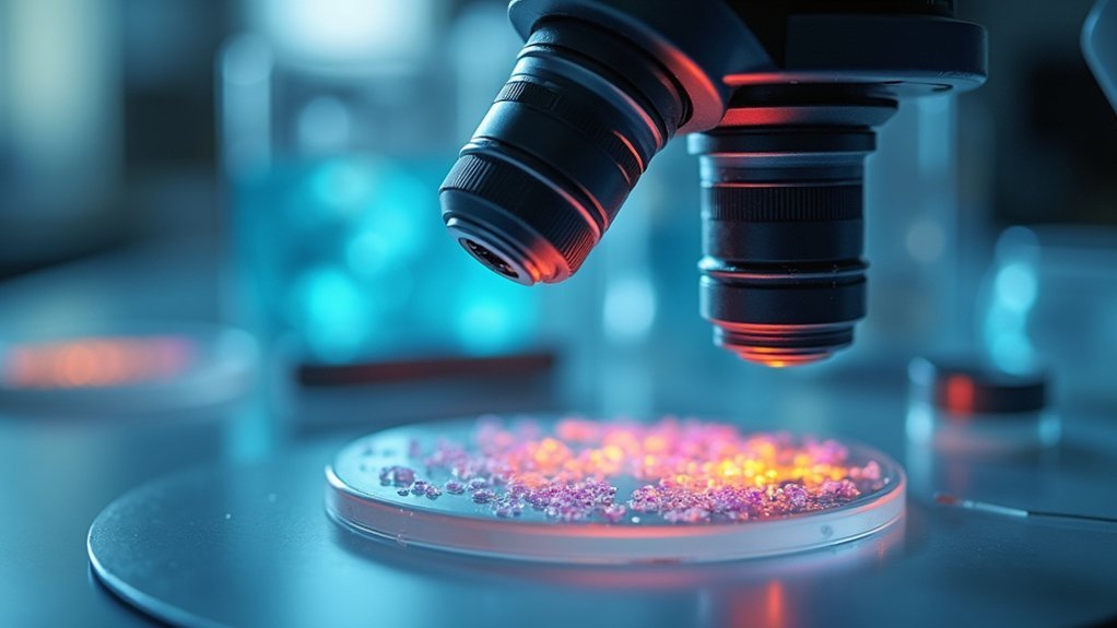
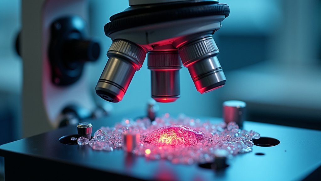
Crystal clear live focus for specimens requires an integrated approach: reliable autofocus systems with stabilization algorithms, controlled environmental conditions, and proper LED illumination. You’ll need regular equipment calibration, anti-vibration tables, and piezoelectric stages for nanometer precision. Consider using AI-driven resolution amplification and noise reduction tools that achieve up to 97% accuracy in detail detection. Mastering both automatic and manual techniques guarantees you’ll capture the dynamic cellular processes that might otherwise remain invisible to the untrained eye.
8 Second-Level Headings for “What Makes Live Focus Crystal Clear For Specimens?”

When you’re capturing dynamic cellular processes, the quality of your focus can make or break your research. Maintaining crystal clear imaging requires understanding and controlling several key factors.
First, you’ll need to implement reliable autofocus systems that can make real-time adjustments during observation. These systems detect and correct focus drift automatically, saving you from blurred images and misinterpreted data.
Second, carefully monitor environmental conditions in your imaging setup. Temperature fluctuations and humidity changes can greatly impact focus stability. Creating a controlled environment minimizes these variables.
Third, establish a regular calibration schedule for your equipment. Even high-end microscopes require consistent maintenance to perform at their best.
Finally, consider using focus stabilization algorithms that compensate for mechanical vibrations and sample thickness variations, ensuring your specimens remain perfectly clear throughout extended imaging sessions.
The Science Behind Live Focus Technology
As researchers explore deeper into cellular dynamics, the sophisticated mechanisms behind live focus technology become increasingly essential for breakthrough discoveries.
When sending us your Lab Work that requires live imaging, you’ll benefit from one of the key innovations in microscopy: piezoelectric stages that provide nanometer-level precision for maintaining perfect focus.
Advanced autofocus systems continuously adjust in real-time to counteract focus drift caused by temperature changes and mechanical vibrations.
Intelligent autofocus technology delivers real-time adjustments, eliminating drift variables for impeccable microscopic visualization.
These systems integrate focus stabilization algorithms that work tirelessly throughout your imaging sessions.
Today’s equipment requires regular calibration to guarantee consistent results during extended observations.
The emerging field of machine learning is revolutionizing this process by predicting necessary focus adjustments before drift occurs, making your specimen visualization more reliable and crystal clear than ever before.
Essential Equipment for Perfect Specimen Clarity

To achieve unparalleled clarity in live specimen imaging, you’ll need to invest in specialized equipment that forms the foundation of reliable microscopy. High-quality optical components with elevated numerical apertures should top your list, as they dramatically enhance resolution and clarity.
You’ll find that implementing autofocus systems and focus stabilization algorithms prevents blur from focus drift during extended imaging sessions. Consider adding a piezoelectric stage to your setup for precise focus control when capturing dynamic cellular processes.
Don’t overlook the importance of regular equipment calibration to maintain consistent image quality.
Finally, mount your entire system on an anti-vibration table to eliminate external disturbances that compromise clarity. This combination of precision components creates an ideal environment for capturing crystal-clear live specimen images.
Optimizing Lighting Conditions for Crystal Clear Results
Your sophisticated microscopy equipment can only perform at its peak when paired with masterful lighting techniques.
LED illumination offers stability while minimizing heat damage to your live specimens, preserving their natural state throughout imaging sessions.
Fine-tune your approach with these critical adjustments:
- Control light intensity to eliminate glare and overexposure that mask essential cellular details.
- Select specific wavelengths like blue or green to enhance visibility of targeted structures and reduce background noise.
- Incorporate diffusers or polarizers to create even illumination across your field of view, eliminating shadows that distort accurate imaging.
Mastering Manual Focus Techniques for Live Specimens

To master manual focus for live specimens, you’ll need to anticipate movement by establishing focus points slightly ahead of the specimen’s predicted path.
Bracketing your focus points—setting multiple focal planes above and below your subject—ensures you’ll capture clear images despite unexpected specimen shifts.
Proper microscope setup, including regular calibration and employing piezoelectric stages, forms the foundation for precise manual focusing and minimizes troublesome focus drift during critical observations.
Anticipate Specimen Movement
Why do even the most carefully prepared live specimens often frustrate microscopists? The answer lies in movement—cellular processes don’t pause for your observations.
Focus drift can transform crisp images into blurry disappointments, potentially compromising your entire experiment.
To stay ahead of specimen movement:
- Establish a manual refocusing schedule – Set consistent intervals to check and adjust focus, especially when observing rapid cellular events.
- Utilize piezoelectric stage technology – These precision tools allow for instantaneous focus adjustments when you detect specimen drift.
- Implement predictive autofocus systems – Modern systems designed for live imaging can anticipate and compensate for movement in real-time.
Remember that equipment calibration and stable environmental conditions minimize focus drift, making your manual techniques more effective during lengthy observation sessions.
Bracketing Focus Points
When live specimens refuse to stay in a single focal plane, bracketing focus points becomes an important technique for capturing clear, usable images. By taking multiple shots at different focus settings, you’ll guarantee at least one image captures ideal clarity for accurate data interpretation.
This approach enhances depth of field in live-cell microscopy, revealing intricate cellular structures that a single focus point might miss. Incorporate autofocus systems to make real-time adjustments as specimens move, maintaining focus during dynamic imaging sessions.
Complement your bracketing technique with manual refocusing at regular intervals to account for variations in sample thickness and refractive index.
Don’t overlook equipment calibration—it’s vital for minimizing focus drift that can compromise image quality. These practices will dramatically improve your ability to document living specimens with precision.
Proper Microscope Setup
The foundation of successful live specimen imaging lies in meticulous microscope setup before you even place your sample on the stage.
Environmental stability is essential—you’ll need to maintain consistent temperature and humidity to prevent focus drift during observation sessions.
Your equipment requires regular calibration to eliminate mechanical instabilities that lead to blurred images.
Consider investing in a piezoelectric stage for nanometer-precise focus adjustments when tracking dynamic cellular processes.
To achieve crystal-clear focus throughout your imaging session:
- Implement an autofocus system or focus stabilization algorithm to maintain clarity during extended observations
- Set a schedule for manual refocusing at predetermined intervals to guarantee reproducibility
- Control environmental conditions rigorously to minimize thermal expansion effects on your optical system
Digital Enhancement Tools for Microscopic Detail

Modern AI-driven resolution amplification technologies can transform your standard microscopic images into remarkably detailed visualizations that reveal previously invisible cellular structures.
You’ll find virtual noise reduction capabilities particularly valuable when working with low-light specimens, as they eliminate background static without compromising critical biological details.
Contrast enhancement algorithms further refine your microscopic observations by automatically adjusting image parameters to highlight subtle differences in tissue density and cellular composition.
AI-Driven Resolution Amplification
Revolutionary AI-driven resolution amplification has transformed how researchers visualize microscopic specimens. These intelligent systems analyze and reconstruct images, giving you markedly clearer views of your specimens without compromising detail integrity.
You’ll notice immediate improvements in your microscopy work with:
- Up to 97% recall rates for crystal detection—far surpassing human capabilities
- Dramatic reduction in noise and artifacts, preserving critical specimen details
- Faster processing times requiring less computational power than traditional methods
Machine learning algorithms adapt to your specific imaging conditions, optimizing focus even in challenging environments.
This means you’ll achieve sharper, more defined visuals regardless of specimen type or lighting variables. With AI enhancement, you’re able to extract meaningful data from images that would otherwise remain obscured by technical limitations.
Virtual Noise Reduction
While focusing on live specimens under a microscope, you’ll often encounter unwanted visual static that obscures critical details. Virtual noise reduction tools solve this problem by deploying sophisticated algorithms that filter out background noise, dramatically enhancing image clarity.
You’ll notice an immediate improvement in signal-to-noise ratio as these digital tools adaptively clean your microscopic images. By leveraging machine learning models, the software analyzes and removes specific noise patterns unique to your data. This smart filtering preserves delicate cellular structures while eliminating distractions.
When you’re studying dynamic cellular processes in real-time, these enhancement techniques help maintain focus and detail throughout your observation period.
You’ll make more accurate interpretations of your microscopic data, ultimately leading to stronger research outcomes and clearer visualization of your specimens.
Contrast Enhancement Algorithms
Because subtle cellular details often hide in plain sight, contrast enhancement algorithms serve as vital tools in your microscopic imaging arsenal.
These powerful digital techniques can transform unclear images by adjusting brightness and contrast levels, making previously invisible features suddenly apparent.
When you’re studying live cells, you’ll benefit from:
- Up to 40% improved clarity through techniques like histogram equalization and adaptive contrast enhancement
- Enhanced differentiation between specimens and backgrounds, important for accurate interpretation
- Significant noise reduction and edge enhancement for better visualization of cellular structures
You’ll find that implementing these digital tools dramatically improves your ability to capture and interpret cellular events and dynamic processes.
This enhancement not only makes your observations more precise but also guarantees your research results remain reproducible and reliable.
Preventing Focus Drift During Extended Observations

Since successful live-cell imaging often requires hours or even days of continuous observation, preventing focus drift becomes critical for maintaining high-quality data collection. You’ll need to implement multiple strategies to combat this common issue.
| Strategy | Benefit |
|---|---|
| Temperature stabilization | Prevents thermal expansion/contraction of optical components |
| Anti-vibration tables | Minimizes mechanical disturbances to the imaging system |
| Autofocus systems | Makes real-time adjustments to maintain clarity |
| Regular calibration | Guarantees focus settings remain accurate over time |
| Piezoelectric stages | Provides precise control for quick drift corrections |
Calibration Strategies for Different Specimen Types
As you work with diverse biological specimens, you’ll need to adopt calibration approaches specifically tailored to each sample type. Different specimens possess unique optical properties that require adjustments to your microscopy equipment for ideal focus.
For consistent, clear imaging across specimen types:
- Match calibration frequency to specimen stability – Live specimens require more frequent recalibration due to temperature-induced focus drift compared to fixed samples.
- Incorporate piezoelectric stages for fine focus adjustments that accommodate varying specimen thicknesses and refractive indices.
- Implement adaptable autofocus systems that recognize different specimen characteristics, reducing manual calibration time while maintaining image clarity.
Don’t forget to regularly check optical alignment and lens cleanliness as part of your calibration protocol, ensuring ideal performance regardless of specimen type.
Frequently Asked Questions
What Makes the Image Clearer on a Microscope?
You’ll get clearer microscope images by maintaining proper focus, using autofocus systems, regularly calibrating equipment, employing piezoelectric stages, and keeping optical components clean. Environmental stability also prevents focus drift during imaging sessions.
How Do You Make a Microscope Clearer?
You’ll achieve clearer microscope images by implementing autofocus systems, regularly calibrating optics, using piezoelectric stages, maintaining stable environmental conditions, and employing focus stabilization algorithms. Clean lenses thoroughly before each use for maximum clarity.
In Summary
You’ll achieve crystal clear live focus for specimens when you’ve mastered both the technical and practical aspects of microscopy. By combining quality equipment, proper lighting, refined focusing techniques, and appropriate digital tools, you’re setting yourself up for success. Remember that consistent calibration and drift prevention strategies aren’t optional—they’re essential for maintaining clarity throughout your observations. Practice these approaches methodically, and your specimen images will showcase remarkable detail.


Leave a Reply