
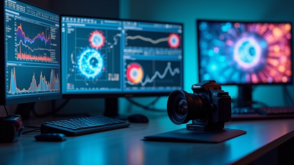
For scientific live capture imaging, you’ll find NINA offers advanced automation while SharpCap excels at real-time stacking. Specialized options like ZEN and NIS-Elements provide AI-powered analysis for professional research, while APT suits beginners with its friendly interface. Your choice depends on specific needs—automation, processing power, or ease of use. Open source alternatives like ImageJ offer customizable solutions for budget-conscious labs. The following comparison reveals which features truly matter for your research applications.
Numeric List of 13 Second-Level Headings
These thirteen scientific imaging software options provide researchers and astrophotographers with specialized tools for capturing, processing, and analyzing celestial objects.
Each package offers unique capabilities to enhance your astronomical imaging workflow.
Specialized astronomical tools crafted to elevate your imaging process from capture to final visualization.
- SharpCap: Real-time live stacking for immediate image quality enhancement
- NINA: Advanced automation including meridian flipping and sequencing
- Voyager: Simplified deep sky object automation with flexible controls
- APT: Beginner-friendly interface with developing auto-focus capabilities
- Maxim DL: All-in-one solution integrating guiding and camera control
- Image Processing algorithms and techniques
- Compatibility with camera hardware
- Guiding capabilities
- Plate solving integration
- User interface comparison
- Automation features
- Cost analysis
- Learning curve assessment
Key Features to Look for in Scientific Imaging Software
When selecting scientific imaging software for astronomical research, you’ll want to focus on specific features that maximize your imaging capabilities. The best Image Analysis Software should offer smart acquisition tools that automate processes and enhance reproducibility.
| Feature Category | What to Look For |
|---|---|
| Automation | Software like ZEISS ZEN with automatic settings generation for various microscope types |
| Processing Tools | Over 180 data transformation options for quantitative, unbiased results |
| Data Handling | GPU-powered 3D rendering capabilities for large datasets (200+ GB) |
Don’t overlook batch processing functionality that streamlines your workflow when analyzing multiple images. Also, make certain the software integrates with third-party platforms, similar to NIS-Elements, creating a unified environment for diverse data acquisition and visualization needs.
Top Microscopy Software Platforms for Live Cell Imaging
You’ll find AI-powered analysis capabilities in top platforms like Aivia and HORIZON™, which transform raw microscopy data into actionable biological insights.
Modern live cell imaging software now offers real-time automated acquisition features, as demonstrated by ZEN’s smart automation and CellReporterXpress’s automated workflows that minimize user intervention.
These automation advances in platforms like NIS-Elements allow you to maintain consistent imaging parameters across experiments while freeing your time for data interpretation rather than repetitive acquisition tasks.
AI-Powered Analysis Solutions
Modern microscopy has been revolutionized by AI-driven software platforms that greatly enhance live cell imaging capabilities. When selecting analysis software, you’ll find several options leveraging deep learning to improve data analysis accuracy and visualization.
| Software | Key AI Feature | Best For |
|---|---|---|
| NIS.ai | Deep learning algorithms | Enhanced accuracy in data analysis |
| Aivia | Real-time processing | Complex live cell imaging scenarios |
| NoviSight 3D | 3D cell model analysis | Drug discovery applications |
| HORIZON™ | Hyperplex image analysis | Detailed biological process insights |
| InVivoAX | Multi-modal algorithms | Advanced live cell applications |
Each platform offers unique AI capabilities that transform raw imaging data into actionable insights. By incorporating these advanced image analysis tools into your workflow, you’ll reveal deeper understanding of cellular dynamics while reducing manual analysis time.
Real-Time Automated Acquisition
Building on advances in AI analysis, real-time automated acquisition software represents the next frontier in microscopy technology. Programs like NIS-Elements and Aivia revolutionize live cell imaging by eliminating manual intervention during critical observation periods, ensuring you’ll never miss key cellular events.
Leica LAS X and ZEN take workflow efficiency to new heights with smart automation features that drastically reduce human input while increasing experimental reproducibility.
For drug discovery applications, NoviSight 3D Cell Analysis Software enables you to work with physiologically relevant 3D models in real time.
When you need to analyze data from multiple imaging modalities, InVivoAX’s cloud-based platform allows side-by-side comparison of diverse datasets.
Meanwhile, NIS.ai enhances your imaging accuracy through deep learning algorithms that continuously improve as you capture cellular behavior.
Automation Capabilities Across Leading Software Solutions
While manual control remains important for specialized imaging situations, automation capabilities have become a defining feature of leading scientific imaging software.
NINA leads the pack with its advanced sequencing customization, creating a user-friendly environment for complex automation tasks during imaging sessions.
SharpCap’s strengths lie in automating real-time processes like polar alignment and image acquisition alongside its live stacking features.
Meanwhile, APT is actively developing automated focusing capabilities to streamline operational workflows.
If you’re seeking balance, Voyager offers simplicity without sacrificing advanced options, making complex imaging task management accessible.
For microscopy applications, ZEN’s smart automation handles hardware calibration across different microscope types, ensuring reproducibility in your acquisition process.
Each solution addresses different automation needs, allowing you to select software that best complements your specific imaging requirements.
Real-Time Processing and Analysis Tools Comparison

Real-time processing capabilities have become essential for scientific imaging applications where immediate feedback drives decision-making. When you’re capturing astronomical image data, your software’s ability to process information instantly can make the difference between a successful observation and a missed opportunity.
| Software | Real-Time Feature | Processing Strength | Best For |
|---|---|---|---|
| SharpCap | Live Stacking | Faint Object Enhancement | Quick Results |
| NINA | Advanced Sequencing | Automation Workflows | Long Sessions |
| Voyager | Dragscript System | Balanced Processing | Flexible Control |
| APT | Developing Auto-Focus | User-Friendly Interface | Beginners |
| Maxim DL | Integrated Guiding | Streamlined Data Flow | All-in-One Use |
Each software offers distinct advantages for processing image data in real-time, allowing you to choose based on your specific scientific imaging requirements and workflow preferences.
Software Integration With Microscope Hardware Systems
Unlike standalone analysis tools, scientific imaging software must maintain seamless communication with microscope hardware to deliver its full potential.
When evaluating software integration capabilities, you’ll find significant differences between major platforms.
ZEISS ZEN provides a universal interface that works across all ZEISS systems, streamlining both routine and complex workflows.
Similarly, Nikon’s NIS-Elements creates a unified environment for data acquisition and visualization with Nikon microscopes.
Leica offers two integrated solutions: LAS X, which facilitates communication between different Leica microscope types, and Aivia, which enables advanced 2-to-5D visualization and analysis.
For specialized applications, Lunaphore’s HORIZON™ demonstrates how tailored software integration with specific hardware (COMET™) can optimize image analysis performance.
Your choice ultimately depends on which manufacturer’s ecosystem you’ve invested in.
Data Management Solutions for Large Image Datasets
As imaging technology advances, scientific researchers face increasingly massive datasets that demand sophisticated management solutions. Modern software like ZEN demonstrates the power of efficient data management solutions by handling datasets up to 200 GB with GPU-powered 3D rendering for ideal visualization.
You’ll find batch processing capabilities essential for analyzing large image collections, providing quantitative and unbiased results without manual intervention. Advanced metadata reading features give you vital context for your images, while lossless compression methods like ZSTD considerably improve file transfer rates and storage efficiency.
When selecting imaging software, look for platforms that integrate with third-party systems, allowing you to work seamlessly with diverse image formats and sources in a unified framework—essential functionality when managing the increasingly complex imaging datasets in today’s research environment.
User Interface and Experience Evaluation
While sophisticated algorithms power scientific imaging software, the user interface ultimately determines how effectively researchers can leverage these capabilities. ZEN Microscopy Software offers a universal interface balancing simplicity for routine work with flexibility for complex research.
You’ll find NINA particularly accessible with its automation features serving both beginners and advanced users in live capture scenarios.
SharpCap prioritizes ease of use during live viewing sessions, especially excelling in its intuitive live stacking functions.
For beginners, APT provides the gentlest learning curve with straightforward setup and operation processes.
In contrast, Maxim DL delivers extensive control by integrating multiple imaging functions into a single screen, though you’ll need to invest more time mastering its interface compared to the other options.
AI-Enhanced Imaging Features in Modern Software

Modern scientific imaging platforms like NIS.ai and ZEN now incorporate AI-driven features that’ll transform your microscopy workflow with intelligent automation and enhanced image processing.
You’ll benefit from real-time data extraction capabilities in software such as HORIZON™ and Aivia, which can immediately analyze complex biological structures while you’re capturing images.
These AI enhancements integrate seamlessly into your research protocols, automating tedious tasks from image acquisition to analysis and allowing you to focus on interpreting results rather than manual processing.
AI-Enhanced Imaging Features in Modern Software
Today’s scientific imaging landscape has been revolutionized by artificial intelligence, with leading platforms integrating sophisticated AI algorithms to enhance both acquisition and analysis capabilities.
You’ll find these AI-enhanced features particularly valuable for processing complex microscopic datasets with unprecedented efficiency.
- ZEN Microscopy Software offers intelligent control mechanisms and smart automation that greatly improve reproducibility across your experiments.
- HORIZON™ software specializes in hyperplex image analysis, giving you advanced visualization capabilities for complex COMET™ datasets.
- Aivia’s AI-driven algorithms streamline your workflow with efficient data processing, supported by 37 academic citations validating its effectiveness.
NIS.ai and NIS-Elements stand out with deep learning techniques that transform how you interpret microscope imaging data, providing analytical power that was unimaginable just a few years ago.
Real-time Data Extraction
As researchers capture increasingly intricate microscopic imagery, real-time data extraction capabilities have become essential for immediate analysis and decision-making. You’ll find that modern software solutions now leverage AI and deep learning to transform how you interact with live microscopy data.
| Software | Real-time Data Extraction Capability |
|---|---|
| NIS.ai | AI-powered algorithms enhance imaging accuracy during live capture |
| Leica LAS X | Intelligent algorithms optimize communication between components |
| NoviSight | Enables monitoring of 3D cell model responses in real-time |
| Aivia | Extracts meaningful data from complex images as they’re captured |
These platforms eliminate the traditional delay between image acquisition and analysis. Whether you’re monitoring drug responses in NoviSight or visualizing complex structures with Aivia, real-time data extraction dramatically improves experimental efficiency and allows for on-the-fly adjustments to your imaging parameters.
Automated Workflow Integration
While capturing high-quality images remains fundamental, the integration of AI-enhanced automated workflows has revolutionized scientific imaging software.
Today’s platforms like NIS.ai and ZEN Microscopy Software leverage deep learning to automate complex analysis processes, dramatically improving efficiency and reducing human error.
- Streamlined processes – Software like Aivia provides over 180 image processing tools for batch processing and quantitative analysis, ensuring unbiased results.
- Multi-modal integration – InVivoAX’s algorithms enable side-by-side analysis of different imaging data types, creating thorough automated workflow integration.
- Smart automation – NoviSight and ZEN software simplify hardware calibration and image acquisition, making reproducible results accessible across different imaging systems.
These AI-driven capabilities transform raw images into actionable data, allowing you to focus on interpretation rather than tedious processing steps.
Cost Analysis and Value Assessment of Premium vs. Free Options
When evaluating scientific imaging software options, researchers must carefully weigh the cost-benefit ratio between premium and free alternatives.
Premium packages like NIS-Elements and Leica LAS X can exceed $10,000 but offer advanced capabilities, dedicated support, and seamless microscope integrations that justify their cost analysis for professional research settings.
While premium imaging solutions command significant investment, their specialized features and robust support infrastructure represent essential value for serious research environments.
Free options such as NINA and SharpCap provide essential functionalities without financial commitment, making them ideal for beginners or budget-conscious labs. They’re praised for ease of use and basic automation features, though they typically lack sophisticated analysis tools and regular updates.
Many premium vendors offer trial versions so you can test their value against free alternatives before investing.
This hands-on assessment helps guarantee you’re selecting software that delivers the specific capabilities your research requires without overpaying for unused features.
Performance Benchmarks for High-Speed Image Acquisition
Beyond cost considerations, the technical capabilities of imaging software greatly impact research outcomes, particularly in time-sensitive applications.
When evaluating high-speed image acquisition tools, you’ll find significant performance variations across platforms.
- SharpCap’s live stacking capability delivers high quality results during real-time observations, dramatically improving collection efficiency.
- NINA’s automation features handle complex sequences and meridian flipping with minimal intervention, essential for extended imaging sessions.
- ZEN by ZEISS processes large datasets rapidly through smart automation and batch processing.
The right software selection depends on your specific needs.
Maxim DL provides extensive control through a unified interface for capturing fast-moving subjects, while Voyager offers an approachable platform suitable for both beginners and experts.
Your research requirements should guide which performance benchmarks matter most.
Multi-Dimensional Imaging Support Across Platforms
As research complexity increases, multi-dimensional imaging has become essential for scientists seeking to capture biological structures in their full depth and context.
When selecting software for your lab, you’ll find varied multi-dimensional imaging support across platforms.
ZEN leads with over 180 image processing tools, enabling efficient data management for complex samples.
ZEN transforms overwhelming data into actionable insights with its comprehensive toolset for demanding multi-dimensional imaging applications.
Leica LAS X excels by integrating various microscope types and offering seamless navigation between data layers.
If you’re working across both 2D and 3D environments, Nikon’s NIS-Elements provides a unified environment specifically designed for microscopy.
For more advanced applications, Aivia’s 2-to-5D visualization capabilities handle intricate microscopic datasets, while Lunaphore’s HORIZON™ specializes in hyperplex image analysis, making complex data interpretation more accessible and efficient.
Open Source Alternatives to Commercial Imaging Software
While commercial platforms dominate many research environments, open source imaging software provides powerful alternatives that won’t strain your budget. ImageJ and its distribution FIJI offer extensive functionality through plugins specifically designed for scientific applications, including 3D visualization and fluorescence microscopy analysis.
- GIMP delivers advanced image editing capabilities comparable to proprietary solutions, making it ideal for post-processing scientific images.
- Micro-Manager gives you direct control over microscopes and imaging devices with automation features that rival commercial counterparts.
- Community-driven development guarantees these tools evolve rapidly to meet specific research needs.
The open source approach fosters collaboration and innovation, creating diverse toolsets that can be customized for your unique imaging requirements—all while maintaining professional-grade quality and compatibility with scientific workflows.
Frequently Asked Questions
How Steep Is the Learning Curve for Beginners?
Your learning curve depends on your technical background. If you’re comfortable with software, you’ll pick it up quickly. Otherwise, expect a few weeks of practice before you’re fully confident maneuvering the features.
Can These Software Options Process Historical Image Datasets?
Yes, you’ll find most options process historical datasets. ImageJ excels with legacy formats, while ZEN and NIS-Elements offer batch processing features. You’ll need to verify specific format compatibility for your archived images.
What Security Features Protect Sensitive Imaging Data?
Security features protecting your sensitive imaging data include encryption, access controls, audit trails, compliance certifications (HIPAA/GDPR), secure cloud storage, and authentication protocols. You’ll want software that offers these protections as standard.
How Frequently Are Software Updates Typically Released?
You’ll typically see software updates every 1-3 months for imaging programs. Major releases come annually, while critical security patches are pushed immediately. Update frequency varies by vendor and product maturity.
Do Any Options Offer Remote Access for Collaboration?
You’ll find several options with remote collaboration features. ZenCapture Pro and BioImager Cloud offer real-time sharing capabilities, while MetaMorph and ImagePro Plus support secure remote access through team licenses and VPN connections.
In Summary
You’ve seen how the right scientific imaging software can transform your live capture workflow. When selecting your solution, prioritize capabilities that match your specific research needs and budget constraints. Whether you opt for commercial powerhouses or open-source alternatives, choose software that balances real-time processing, automation, and multi-dimensional support. Your imaging strategy directly impacts your research outcomes—invest wisely in tools that’ll enhance your scientific discoveries.


Leave a Reply