
For professional research, consider Leica STELLIS STED for super-resolution clarity or Nikon’s multichannel fluorescence systems for in vivo applications. If you’re budget-conscious, the Celestron CM800 ($87) offers 800x magnification, while Bresser Biolux NV ($274) includes camera capabilities. Don’t overlook software platforms like Leica MICA that streamline workflows. Pairing your system with environmental control chambers will dramatically improve experimental consistency and research outcomes.
Understanding the Essential Features of Live Cell Imaging Systems
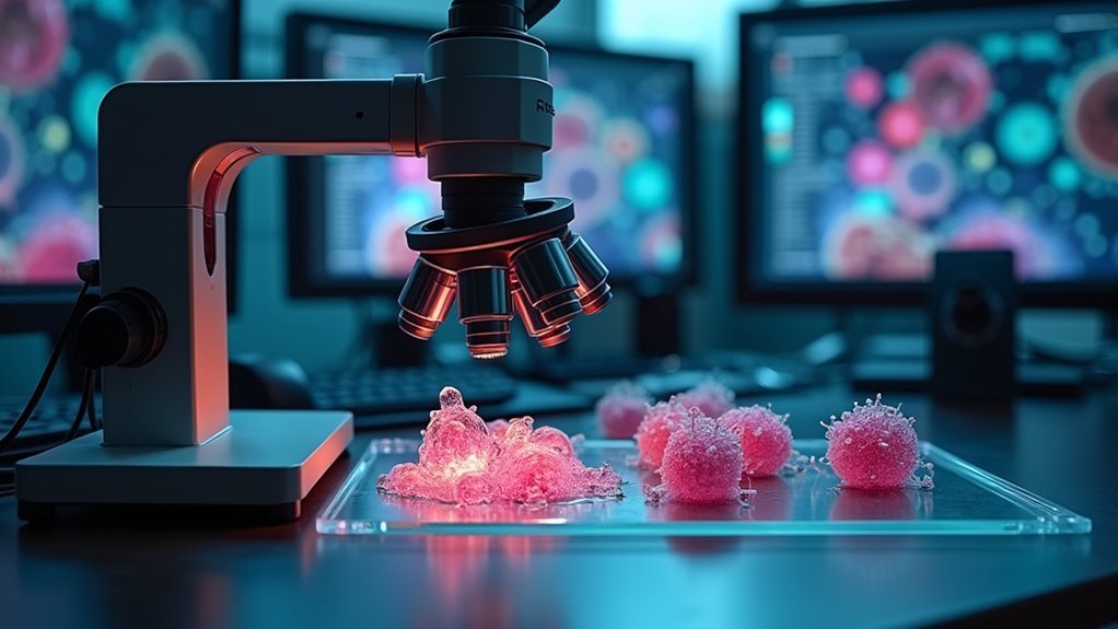
Innovation in cellular research hinges on your ability to observe living cells in real time. When selecting a live cell imaging system, you’ll need to prioritize features that align with your specific research goals.
The cornerstone of cellular discovery lies in real-time observation, demanding imaging systems precisely matched to your research objectives.
Look for systems offering high-magnification capabilities and compatibility with various imaging techniques such as fluorescence, confocal, and super-resolution microscopy. These technologies reveal cellular dynamics and interactions with remarkable detail.
Your ideal system should support advanced applications like FRET and FRAP for specialized studies.
User-friendly software is equally important, as it streamlines data acquisition and analysis, saving you valuable research time. Top manufacturers like Molecular Devices and Leica Microsystems provide reliable options that balance sophisticated functionality with intuitive operation.
The right imaging system will serve as your window into the microscopic world of cellular processes.
Top Professional-Grade Microscope Systems for Research Laboratories
Five leading microscope systems distinguish themselves in today’s research landscape, each offering specialized capabilities for advanced live cell imaging.
The Leica STELLARIS STED delivers super-resolution clarity for detailed cellular processes, while Nikon’s multichannel fluorescence systems excel at real-time in vivo applications.
For research labs seeking user-friendly operation without sacrificing quality, Keyence’s BZ-X800 provides advanced imaging technology specifically designed for biological research.
If your focus is cell line development, consider Molecular Devices’ CloneSelect Imager, which precisely assesses cell viability during live imaging sessions.
When working with larger specimens, Miltenyi Biotec’s UltraMicroscope II stands out as your best option, providing exceptional fluorescence imaging for whole organisms or substantial tissue samples within a thorough imaging system.
Budget-Friendly Live Imaging Solutions for Educational Settings
While advanced research laboratories require cutting-edge microscope systems, educational institutions can achieve excellent results with more affordable alternatives.
The Celestron CM800 Compound Microscope offers impressive 800x magnification for just $87.19, delivering excellent optical quality without breaking the bank.
For younger students, consider the National Geographic Dual LED Microscope at $67.99, providing 20x-50x magnification for basic specimen exploration.
If you need more versatility, the Bresser Biolux NV ($274.53) includes built-in camera capabilities for live imaging across diverse educational applications.
The ultra-portable Celestron S20 ($49.67) is perfect for field trips with its lightweight design and built-in LED illumination.
While budget brands like Amscope and Omax exist, mid-range options like the CM800 offer better reliability for serious educational settings.
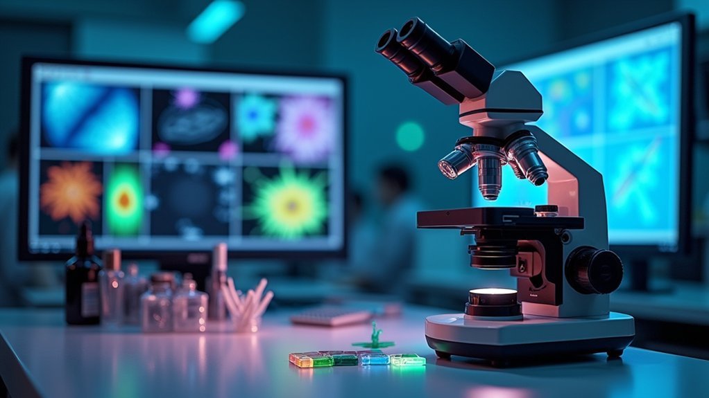
Advanced Software Capabilities for Real-Time Cellular Analysis
Moving beyond educational solutions, today’s advanced microscope systems incorporate powerful software that transforms passive observation into active analysis.
You’ll find sophisticated platforms like Leica MICA and Nikon Ji offering user-friendly interfaces that streamline your workflow while delivering powerful analytical capabilities.
These advanced software packages enable you to:
- Watch cellular migration and division as they happen, with real-time quantitative metrics appearing on your screen
- Leverage specialized techniques like FRET and FRAP to visualize molecular interactions at the cellular level
- Immediately assess experimental results without waiting for post-processing, accelerating your research timeline
With real-time cellular analysis capabilities, you’ll make faster discoveries while maintaining experimental reproducibility and data integrity—essential advantages in competitive fields like drug development and cellular biology research.
Accessories That Enhance Your Live Microscopy Experience
Environmental control chambers maintain your cells’ ideal temperature, pH, and humidity conditions, ensuring they remain viable during extended imaging sessions.
Digital recording solutions from leading manufacturers let you capture every critical cellular event in real-time with precise timing and minimal photobleaching.
Specialized sample holders complement these accessories by stabilizing your specimens and allowing for multi-position imaging across various tissue samples or cell cultures.
Environmental Control Chambers
A stable microenvironment forms the cornerstone of successful live cell imaging. Environmental control chambers regulate temperature, humidity, and CO2 levels to mimic cells’ natural conditions, ensuring your specimens remain viable during extended observation periods.
By incorporating these chambers into your imaging setup, you’ll capture more accurate cellular processes and responses without stress-induced artifacts.
- Visualize healthy cells dividing naturally under perfect physiological conditions for hours or even days
- Picture seamlessly switching between imaging modalities while maintaining stable environmental parameters
- Imagine collecting reliable data that truly represents in vivo cellular behavior
These chambers integrate with major systems from manufacturers like Leica and Nikon, making them essential investments for any serious live cell imaging work.
You’ll see dramatic improvements in experimental outcomes when cells aren’t struggling against environmental fluctuations.
Digital Recording Solutions
While maintaining ideal environmental conditions preserves cell viability, capturing those cellular events requires powerful digital recording capabilities. Modern microscopes integrate seamlessly with USB digital cameras, allowing you to document high-quality images and videos of your live specimens.
Systems like KEYENCE’s BZ-X800 Fluorescence Microscope offer real-time data acquisition features that enhance your analysis workflow. You’ll appreciate how high-resolution cameras reveal fine cellular details that might otherwise go unnoticed during experiments.
The software accompanying these solutions provides valuable tools—image stitching, automated analysis, and quantification—that streamline your research process.
Specialized Sample Holders
Maintaining stable, precise positioning of your specimens represents one of the most critical factors in successful live-cell imaging experiments.
When paired with quality lenses, specialized sample holders dramatically enhance your data quality by minimizing motion artifacts and providing consistent imaging conditions.
Consider these game-changing accessories:
- Molecular Devices LLC holders deliver ideal stability for cell cultures, allowing you to capture cellular processes without disruption.
- Miltenyi Biotec’s UltraMicroscope II holders excel with larger specimens, enhancing fluorescence imaging capabilities.
- Keyence Corporation’s BZ-X800 holders accommodate diverse biological applications with precise positioning features.
These customizable solutions improve both accessibility and ease of use for your imaging systems.
You’ll find it considerably easier to conduct real-time observations while minimizing sample disturbance—ultimately leading to more reliable results in your dynamic cellular studies.
How to Choose the Right System Based on Application Requirements
Selecting the ideal microscope live imaging system begins with thoroughly evaluating your specific research needs. When reviewing application requirements, consider whether you’ll need fluorescence capabilities for cell viability studies or confocal options for detailed drug discovery work. Image quality requirements will vary based on whether you’re studying rapid cellular processes that demand FRET or FRAP techniques.
| Application Type | Recommended Features | Example Systems |
|---|---|---|
| Cell Viability | Basic fluorescence | Olympus APX-100 |
| Drug Discovery | High-resolution | Leica MICA |
| Cellular Process | FRET/FRAP capability | Zeiss LSM |
Don’t overlook software interface usability and future upgrade potential when making your selection. User-friendly systems will enhance your workflow efficiency, while expandable platforms guarantee your investment remains relevant as your research evolves.
Frequently Asked Questions
What Is the Best Microscope for Live Cells?
For live cell imaging, you’ll find the Leica STELLARIS STED offers superior resolution, while KEYENCE’s BZ-X800 provides versatility. Your choice depends on whether you need super-resolution or multi-technique capabilities for your specific application.
What Microscope Is Best for Living Things?
For observing living things, you’ll find the Leica STELLARIS STED excellent for real-time cellular studies, while Nikon’s systems offer versatile in vivo imaging. The UltraMicroscope II is ideal for larger living specimens.
Which Company Is Best for Microscopes?
There’s no single “best” company for microscopes. Your ideal choice depends on your specific needs – Nikon and Leica excel for research, KEYENCE for industrial applications, and Molecular Devices for cell-based work.
What Can You See With a 1000X Microscope?
At 1000x magnification, you’ll see cellular structures like nuclei, mitochondria, and cell membranes. You can observe bacteria, protozoa, tissue sections, and watch live cellular processes such as cell division and movement.
In Summary
You’ve now explored the best live imaging microscopy systems across various price points and applications. Whether you’re conducting cutting-edge research or teaching basic cell biology, the right system combines optical quality, software capabilities, and appropriate accessories. Don’t overlook your specific requirements when making your selection—invest in technology that’ll grow with your needs and deliver reliable, high-quality imaging for years to come.


Leave a Reply