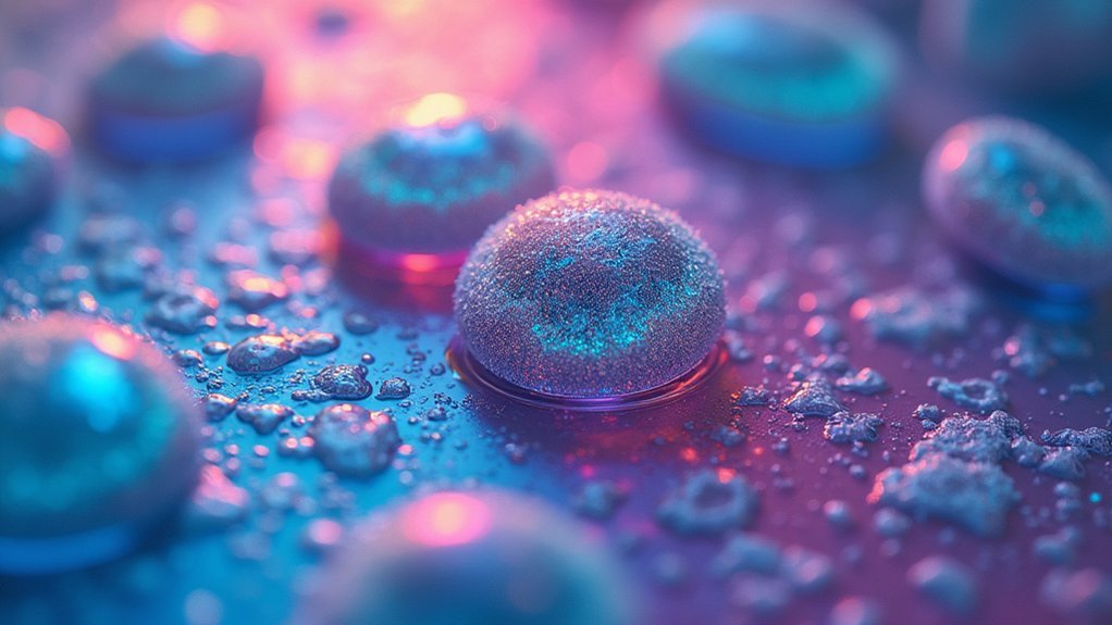
Oil immersion lenses dramatically improve cell image quality by matching the refractive index between your lens and specimen. This eliminates light scattering and reflections that occur with dry objectives, increasing the numerical aperture to 1.3-1.7 (compared to air’s maximum of 1.0). You’ll observe cellular structures as small as 1-2 micrometers with enhanced brightness, contrast, and resolution. These specialized objectives reveal essential details like nuclear pores and mitochondrial cristae that remain invisible with standard lenses.
Second-Level Headings for Blog Post: “Why Oil Immersion Lenses Create Superior Cell Images”

When planning your blog post about oil immersion lenses, you’ll need clear, informative headings that guide readers through the technical advantages these specialized tools offer.
Consider these second-level headings to structure your content effectively:
- “The Science Behind Oil Immersion: Refractive Index Explained”
- “Breaking Resolution Barriers: How Oil Immersion Objectives Enhance Detail”
- “Numerical Aperture: Why Higher NA Values Matter for Cell Imaging”
- “From Invisible to Visible: Capturing Structures Under 2 Micrometers”
- “Applications in Cell Biology: When to Use Oil Immersion Techniques”
- “Practical Tips: Optimizing Your Microscope for High Resolution Imaging”
These headings follow a logical progression from theory to practice, helping your readers understand both the scientific principles and practical applications of oil immersion microscopy.
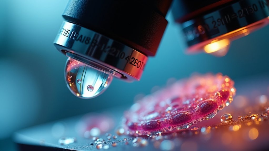
The Physics Behind Oil Immersion Microscopy
When you look through an oil immersion lens, you’re witnessing the magic of refractive index matching, where the oil (n≈1.51) creates a nearly seamless optical path between specimen and objective.
This continuous medium allows more light to enter the objective, boosting the numerical aperture up to 1.7—far above the 1.0 ceiling of dry objectives.
The increased light capture directly enhances your resolution according to the Rayleigh Criterion, letting you see cellular details as small as 1-2 micrometers that would otherwise remain invisible.
Refractive Index Principles
At the heart of oil immersion microscopy lies a fundamental physical property known as refractive index (RI). When you use immersion oil with an RI of about 1.51—similar to glass—you’re creating an ideal pathway for light to travel from your specimen to the objective lens.
| Medium | Refractive Index | Effect on Imaging |
|---|---|---|
| Air | 1.0 | Limited NA (≤1.0), light loss |
| Water | 1.33 | Improved NA, some refraction |
| Immersion Oil | 1.51 | Maximum NA (up to 1.7), minimal light loss |
Without oil, light bends dramatically when moving between the glass slide and air, causing significant loss of detail. By matching the RI of your imaging system components, you’ll capture more light, achieve higher numerical aperture values, and dramatically improve image quality and resolution of cellular structures.
Light Capture Mechanics
The physics of light behavior explains why oil immersion creates such superior cellular images. When you place immersion oil between your specimen and objective lens, you’re creating an ideal pathway for light transmission that dramatically improves resolution.
- Oil eliminates air gaps by matching the refractive index of glass, minimizing light refraction at boundaries.
- Your microscope achieves numerical apertures of 1.3-1.7 with oil immersion (significantly higher than air objectives).
- You’ll capture more light with oil immersion, producing brighter images essential for detailed cellular analysis.
- The Rayleigh Criterion equation shows that higher NA values from oil immersion allow you to resolve structures as small as 1-2 micrometers.
This enhanced light capture is particularly valuable when you’re working at high magnifications like 60x or 100x.
Resolution Enhancement Factors
Resolution enhancement factors explain precisely why oil immersion microscopy revolutionizes cellular imaging capabilities.
When you place immersion oil between your objective lens and specimen, you’re leveraging fundamental physics to overcome traditional limitations.
The primary advantage stems from the oil’s refractive index (n ≈ 1.51), which closely matches glass. This similarity minimizes light refraction at interfaces, allowing more light to reach your detector.
According to the Rayleigh Criterion, resolution improves as numerical aperture increases – oil immersion lenses achieve remarkable NA values of 1.3-1.7 compared to air objectives’ maximum of 1.0.
This enhancement isn’t trivial – you’ll resolve structures as small as 1-2 micrometers that would otherwise blur together.
For high-magnification work (60x-100x), oil immersion becomes essential for distinguishing fine cellular details that air objectives simply can’t resolve.
How Refractive Index Enhances Image Resolution
When examining cellular structures at the microscopic level, refractive index plays an essential role in determining image quality and resolution. Oil immersion creates a continuous optical path between your specimen and objective lens by matching the refractive index of glass (approximately 1.51), which greatly enhances image clarity.
- You’ll capture more light with oil immersion, as the matched indices prevent light loss at interfaces.
- Resolution of the image improves considerably due to reduced diffraction effects.
- High NA values (up to 1.7) become possible, allowing you to distinguish structures as small as 1-2 micrometers.
- Your cellular organelles appear sharper because the oil eliminates refractive distortions that would normally blur fine details.
Comparing Air vs. Oil Immersion Objectives in Cellular Imaging

Although both types serve important roles in microscopy, air and oil immersion objectives differ dramatically in their ability to reveal cellular details. When you’re examining cellular structures, the numerical aperture becomes vital—oil immersion lenses deliver NA values of 1.3-1.7 compared to air objectives’ maximum of 1.0.
This difference exists because immersion oil eliminates light refraction at the lens-sample interface by matching glass’s refractive index (n=1.51). The result? You’ll capture considerably more light with oil objectives.
At higher magnification (60x-100x), oil immersion provides superior resolving power, distinguishing structures as small as 1-2 micrometers that air objectives simply can’t visualize.
While oil objectives require more maintenance to prevent dried oil buildup, their capabilities make them indispensable for microbiology and single-cell analysis.
Critical Cellular Structures Only Visible With Oil Immersion
Many cellular structures remain hidden from view until you employ oil immersion objectives with their superior resolving capabilities.
When you apply immersion oil between your slide and objective lens, you’ll reveal visualization of cellular components as small as 1-2 micrometers that require higher resolution for clear imaging.
With high magnification enhanced by oil immersion, you’ll observe:
- Nuclear pores and chromatin arrangements that reveal cellular regulatory mechanisms
- Mitochondrial cristae and fine structural details essential for metabolic studies
- Ribosomal clusters and endoplasmic reticulum networks that appear sharper and more defined
- Fluorescently labeled nucleic acids during FISH techniques with exceptional clarity
The numerical apertures of 1.3-1.7 achieved with immersion oil greatly improve your ability to monitor live cell dynamics and intracellular processes in real-time.
Numerical Aperture and Its Impact on Image Quality
Numerical aperture stands as the single most important specification for microscope objectives, directly affecting your image’s resolution and clarity.
Numerical aperture determines how much microscopic detail you can capture—the higher the NA, the sharper your image.
When you use oil immersion lenses, you’ll achieve NA values of 1.3-1.7, far exceeding air objectives’ theoretical maximum of 1.0.
This higher NA dramatically improves your image quality by allowing you to distinguish structures mere nanometers apart. The formula NA = n × sin(μ) explains why immersion oil works so effectively—its refractive index (n) is similar to glass, minimizing light refraction at the specimen-lens interface.
For visualizing tiny cellular structures like organelles or bacteria (1-2 micrometers), this enhanced resolution is critical. You’ll capture fine details that would remain invisible with standard dry objectives, making oil immersion essential for serious cellular imaging work.
Selecting the Right Immersion Oil for Different Cell Types
You’ll need to match your immersion oil’s refractive index closely to that of glass (approximately 1.51) to prevent light scattering and enhance cellular detail resolution.
When working with living specimens, consider using lower viscosity synthetic oils that allow easier manipulation while maintaining optical clarity and reducing potential toxicity to cells.
Remember that different microscopy techniques may require specific oil formulations, so you should select an oil that’s compatible with both your cell type and imaging method.
Viscosity for Living Specimens
When observing living specimens under a microscope, selecting the appropriate immersion oil viscosity becomes critical to maintaining cellular integrity and natural behavior.
You’ll need to balance ideal imaging quality with minimal interference of cellular processes.
- Choose synthetic oils with lower viscosity for live cell imaging to allow natural movement and prevent mechanical stress.
- Match the refractive index (typically 1.51) of your immersion oil to both glass and specimen for superior light transmission.
- Consider how different cell types respond—motile bacteria and mammalian cultures typically require oils that combine low viscosity with stability.
- Remember that higher purity immersion oils can greatly enhance visualization of fine cellular structures.
The right viscosity guarantees you’ll capture high-resolution images while keeping your living specimens behaving naturally throughout the observation period.
Refractive Index Matching
Beyond viscosity considerations, the refractive index of your immersion oil represents the cornerstone of high-quality cellular imaging. When you select an oil with a refractive index close to 1.51, you’re effectively matching the glass lens properties, minimizing light refraction and enhancing image clarity.
For different cell types, your choice of immersion oil should align with the specimen’s thickness and composition. Synthetic oils offer stable refractive indices and pH levels, essential for consistent results across various cellular structures.
You’ll achieve superior resolution when the oil’s refractive index closely matches your specific specimen. This careful matching not only improves brightness and resolution but also reduces artifacts from mismatched refractive indices.
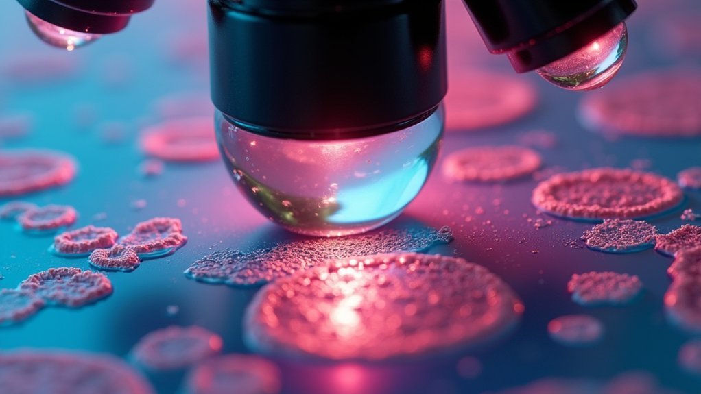
Advanced Techniques in Oil Immersion Microscopy
Although traditional microscopy provides valuable cellular insights, advanced oil immersion techniques have revolutionized how we visualize microscopic structures. When you apply immersion oil with a refractive index similar to glass, you’ll achieve numerical aperture values between 1.3 and 1.7, resolving details as small as 1-2 micrometers.
These enhancements support sophisticated applications:
- Super-resolution imaging – Breaking diffraction limits for nanoscale observations
- Fluorescence in situ hybridization (FISH) – Detecting specific DNA sequences with enhanced contrast
- Live cell imaging – Observing dynamic cellular processes with minimal aberration
- Automated systems like WiScan® Hermes – Precisely applying oil for high-throughput applications
You’ll find these advanced techniques particularly valuable when examining organelles and other subcellular structures that remain invisible with standard objectives.
Common Challenges and Troubleshooting Oil Immersion Systems
You’ll face two common issues when working with oil immersion systems: spillage and autofocus problems.
To prevent oil spillage, always use minimal oil drops and keep absorbent materials nearby during slide changes.
When your microscope struggles with autofocus, check for excessive oil application or contamination, as incorrect oil quantities can interfere with the system’s ability to find the best focal plane.
Oil Spillage Prevention
While using oil immersion lenses provides superior resolution for cellular imaging, preventing oil spillage remains a significant challenge for microscopists.
When working with an oil immersion lens, you must take precautions to avoid contamination of slides and adjacent equipment.
To effectively prevent oil spillage during your microscopy sessions:
- Use automated systems like WiScan® Hermes that apply oil precisely and consistently without manual handling
- Select immersion oil with the appropriate viscosity and refractive index to match your specific lens and slide materials
- Implement regular maintenance and calibration of your oil immersion systems to guarantee peak performance
- Educate yourself on proper application techniques and immediately clean lenses after use
These preventative measures will help maintain image quality and extend the lifespan of your valuable microscopy equipment.
Autofocus Adjustment Issues
Beyond proper oil handling lie the complexities of autofocus systems when working with oil immersion objectives. You’ll encounter challenges when refractive indices between immersion oil and specimens don’t precisely match, disrupting your imaging accuracy.
| Issue | Cause | Solution |
|---|---|---|
| Inaccurate focusing | Refractive index mismatch | Use manufacturer-recommended oil |
| Image distortion | Air bubbles in oil | Apply oil without bubbles |
| Autofocus failure | Mixed oil types | Clean thoroughly between oils |
| Light scattering | Residual oil buildup | Regular lens maintenance |
When your automated system struggles, you may need manual intervention. Troubleshooting typically requires recalibration to account for the specific optical properties of your immersion oil. Remember that proper maintenance, including thorough cleaning after each session, guarantees consistent autofocus adjustment performance and prevents frustrating interruptions during critical imaging work.
Specialized Applications in Microbiology and Pathology
When examining the microscopic world of pathogens and tissue samples, oil immersion lenses become indispensable tools for microbiologists and pathologists alike.
With immersion oil bridging the gap between your objective and specimen, you’ll achieve the high magnification (60x-100x) necessary to visualize bacteria just 1-2 micrometers in size. The increased numerical aperture dramatically improves resolution of critical cellular structures.
Your diagnostic work benefits from oil immersion in four key ways:
- Enhanced visualization of bacterial morphology for accurate identification
- Superior resolution of cellular abnormalities in tissue samples
- Improved brightness and clarity for distinguishing fine structural details
- Better light collection in fluorescence applications for tracking specific cellular markers
These advantages make oil immersion microscopy essential for both static diagnostic work and dynamic live-cell imaging studies.
Historical Development of Oil Immersion Technology
The remarkable capabilities of today’s oil immersion microscopy have their roots in the scientific curiosity of 19th-century microscopists. You’ll find that Ernst Abbe’s groundbreaking 1873 work established the essential relationship between numerical aperture and resolution, revolutionizing microscopy’s potential.
| Era | Innovation | Impact |
|---|---|---|
| Mid-19th century | Concept origin | Reduced light refraction |
| 1873 | Abbe’s formula | Linked numerical aperture to resolution |
| Late 19th century | Commercial lenses | Enabled visualization of smaller structures |
| Early 20th century | Apochromatic objectives | Corrected optical aberrations |
| Late 20th century | Synthetic oils | Stabilized refractive index values |
As you use modern immersion objectives, you’re benefiting from over 150 years of optical refinement that systematically overcame the physical limitations of light microscopy through specialized immersion media.
Digital Imaging With Oil Immersion Objectives
Modern digital imaging has been revolutionized by oil immersion technology, allowing you to capture cellular details previously invisible to conventional microscopy. These objectives achieve higher numerical aperture values up to 1.7, dramatically improving your ability to resolve structures just 1-2 micrometers in size.
Discover extraordinary cell details with oil immersion technology—pushing resolution boundaries for structures once beyond microscopy’s reach.
When you’re capturing digital images of cells, oil immersion objectives offer significant advantages:
- Minimizes light refraction by matching the refractive index (n=1.51) between glass and oil
- Produces brighter, clearer images with enhanced resolution and contrast
- Reveals fine structural details essential for microbiology and cellular studies
- Supports advanced applications like live-cell assays and super-resolution techniques
Automated systems like WiScan® Hermes now facilitate consistent oil application, ensuring you’ll achieve ideal imaging conditions with minimal intervention.
Maintenance and Care for Optimal Performance
Proper maintenance of your oil immersion lenses guarantees their longevity and consistent imaging quality.
Clean your objectives immediately after use to prevent oil from drying and becoming difficult to remove. Use lens paper in a spiral motion from center outward to effectively eliminate residues without scratching the delicate glass surface.
For persistent immersion oil buildup, apply a gentle solution of dishwashing liquid and distilled water, but avoid harsh chemicals that could damage protective coatings.
Store your lenses in dust-free environments and inspect them regularly for any signs of wear. This vigilant care assures your equipment will continue producing high-quality images without degradation.
Remember that properly maintained oil immersion objectives not only perform better but also represent a protected investment in your microscopy work.
Frequently Asked Questions
How Does Immersion Oil Increase Resolution of an Image?
Immersion oil increases resolution by matching glass’s refractive index, which reduces light refraction. You’ll see finer details because it increases numerical aperture, allowing your lens to capture more light with less diffraction.
Why Oil Immersion Objective Lens Is Superior to Dry Objective Lens?
Oil immersion objectives are superior because they’ll give you higher numerical aperture (up to 1.7 vs 1.0), eliminating air-glass refraction that causes light loss. You’ll see finer details with better clarity and brightness.
What Does Oil Do on an Oil Immersion Lens in Relation to the Image?
Oil creates a continuous optical path between your specimen and lens, reducing light refraction and loss. You’ll see brighter, sharper images with higher resolution as more light reaches the objective with minimal distortion.
What Is the Purpose of Using Immersion Oil When Viewing Bacterial Cells With the 100X Objective?
You’ll use immersion oil with 100x objectives to improve resolution and clarity when viewing bacteria. It matches glass’s refractive index, increases numerical aperture, and enhances light collection for visualizing tiny bacterial structures (0.5-5 micrometers).
In Summary
You’ve seen how oil immersion transforms your cellular imaging capabilities by conquering light refraction and maximizing resolution. Whether you’re studying bacterial pathogens or complex tissue structures, these specialized objectives reveal details that would otherwise remain invisible. As you continue your microscopy work, remember that proper care of your oil immersion lenses guarantees they’ll deliver exceptional performance for years to come. The extra effort is always worth the superior results.


Leave a Reply