
Exposure compensation is essential in microscope photography because it helps you capture accurate specimen details that might otherwise be lost in overexposed highlights or underexposed shadows. You’ll need it to overcome the camera’s metering limitations at high magnifications, preserve fine textures, and adapt to varying stain densities. When working with transparent specimens or fluorescent samples, proper exposure adjustments guarantee scientific integrity by maintaining true color representation and revealing authentic structural details. The right compensation techniques can transform your microscopic imaging results.
10 Second-Level Headings for “Why Is Exposure Compensation Crucial In Microscope Photography?”
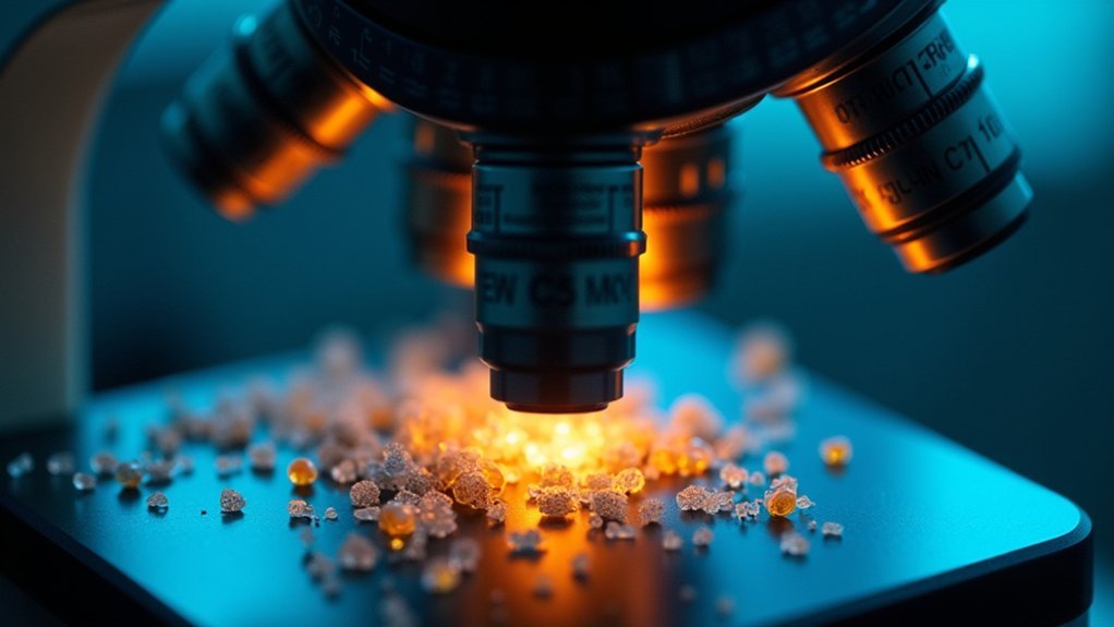
Precision sits at the heart of effective microscope photography, with exposure compensation serving as an essential tool for capturing accurate specimen details.
When you’re examining specimens under high magnification, lighting conditions can vary dramatically across your field of view. Without proper exposure adjustment, you’ll lose vital information in overexposed highlights or underexposed shadows.
Your microscope images need to accurately represent the specimen’s true appearance, which exposure compensation helps accomplish. By making deliberate adjustments to counteract challenging lighting situations, you’ll preserve fine textures and intricate structures that might otherwise be lost.
Precise exposure adjustments reveal the authentic reality of specimens, preserving critical details that define scientific truth.
This becomes particularly important when working with specimens that have both bright and dark regions. Mastering exposure compensation allows you to maintain detail across the entire image, ensuring your scientific documentation remains faithful to what you’re actually observing through the eyepiece.
The Basics of Light Metering in Microscope Photography
When working with microscope photography, you’ll encounter both incident light (directly from your light source) and reflected light (bounced back from your specimen), each requiring different metering approaches.
Your camera’s built-in metering system often struggles with microscope settings because it’s designed for everyday photography rather than the extreme magnification and unusual light paths found in microscopy.
Understanding these limitations helps you recognize when to apply exposure compensation, especially when your specimen creates challenging lighting conditions that confuse automatic metering.
Incident vs. Reflected Light
Although both play essential roles in photography, incident and reflected light metering represent fundamentally different approaches to achieving proper exposure in microscope photography. When you’re working with transparent specimens, incident light measurement provides more consistent results by measuring light falling onto your subject rather than bouncing off it.
| Metering Type | Best For | Challenges |
|---|---|---|
| Incident | Transparent specimens, consistent exposures | Requires separate meter or calibration |
| Reflected | Convenience, built into cameras | Often requires exposure compensation |
| Hybrid | Complex specimens with varying opacity | Demands experience and testing |
Reflected light metering can be misleading with microscope subjects, especially with highly reflective or unusually dark specimens. You’ll often need to apply exposure compensation to correct for these variances, adjusting settings to account for how your specimen’s characteristics affect light readings.
Metering System Limitations
Even the most advanced metering systems have inherent constraints that become starkly apparent in microscope photography. Your camera’s meter is calibrated for standard photography scenarios, not the unique high-contrast environments found under microscopes.
When working with specimens, you’ll notice how meters struggle with accurate readings in high-contrast scenes, often leading to underexposed or overexposed images. The problem worsens with long exposures where reciprocity failure causes the meter to miscalculate necessary exposure settings.
Additionally, your camera’s limited dynamic range fails to capture both bright highlights and deep shadows simultaneously, missing vital specimen details.
This is where exposure compensation becomes essential—it allows you to override these metering inaccuracies and take control of your exposure settings, ensuring your microscopic subjects are properly illuminated and detailed.
Overcoming High Contrast in Specimen Imaging
When imaging microscopic specimens with both bright and dark regions, you’ll often need to apply exposure compensation to capture the full range of details.
You can adjust your exposure by +1/3 to +2/3 EV to prevent highlight clipping while still retaining shadow information in semi-transparent structures.
This balanced approach guarantees you’re preserving the integrity of fine cellular textures and structural elements that might otherwise be lost in high-contrast situations.
Balancing Light-Dark Regions
Microscope specimens with dramatic tonal variations present a unique challenge for photographers seeking accurate representation. When your sample contains both bright and dark areas, your camera’s automatic metering often gets confused, sacrificing detail in shadows or highlights.
Proper exposure compensation helps you overcome these limitations to capture the complete tonal range.
When working with high-contrast specimens, consider these approaches:
- Apply positive exposure compensation (+0.3 to +1.0) to brighten the image when dark areas contain essential details that would otherwise be lost.
- Use spot metering on mid-toned areas of your specimen, then adjust exposure compensation accordingly.
- Try bracketing exposures (shooting multiple images at different compensation values) when dealing with particularly challenging specimens like thick tissue sections.
Preserving Detail Integrity
High-contrast specimens present one of the most significant challenges in microscope photography, threatening the very detail you’re trying to document.
When your camera’s metering system encounters these difficult lighting scenarios, it often misinterprets the proper exposure, causing shadows to darken or highlights to blow out.
By using exposure compensation, you’ll maintain clarity in both bright and dark regions of your specimen.
This is especially critical when working with transparent or translucent samples that unevenly transmit light. You’ll need to adjust the exposure settings to reveal subtle textures and colors that would otherwise be lost.
Understanding characteristic curves will help you fine-tune these adjustments precisely.
When you properly compensate for high contrast, you’re not just creating a better-looking image—you’re preserving the scientific integrity of your documentation.
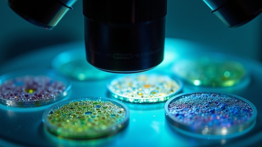
Adapting to Different Staining Techniques
Because various staining techniques dramatically alter specimen light absorption properties, you’ll need to master exposure compensation to capture accurate microscopic images.
Mastering exposure compensation is essential when different stains alter how specimens absorb light under the microscope.
When working with high-contrast stains like hematoxylin and eosin, your camera’s automatic settings may miscalculate proper exposure, requiring manual adjustments to preserve critical details.
For best results with different staining methods:
- Adjust exposure compensation based on stain intensity—immunofluorescence techniques introduce additional light sources that need careful balancing.
- Evaluate specimen thickness before shooting—thicker samples reduce light transmission and often require positive exposure compensation.
- Account for staining uniformity—uneven staining creates varying light reflection patterns that standard metering can’t properly interpret.
Compensating for Fluorescence Microscopy Challenges
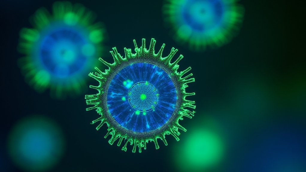
Fluorescence microscopy presents unique exposure challenges that you’ll need to address through targeted compensation techniques.
You can control background signal by adjusting exposure settings to enhance contrast between your specimen’s fluorescent structures and unwanted noise, while implementing strategic interval timing helps mitigate photobleaching that occurs during prolonged light exposure.
To optimize dynamic range, you should bracket your exposures to capture both dim features and bright signals within the same field of view, ensuring thorough data collection from your fluorescent specimens.
Background Signal Control
When working with fluorescence microscopy, you’ll often face the challenge of unwanted background signals that can mask your specimen’s actual fluorescence.
Effective exposure compensation is your key tool for reducing these interfering elements while maintaining the clarity of your target signals.
Background signals can often exceed the intensity of your actual specimen fluorescence, requiring precise adjustment to capture meaningful images.
Here’s how to control these unwanted signals:
- Apply negative exposure compensation when background levels are high to prevent overexposure.
- Fine-tune your settings based on your specific staining method and sample characteristics.
- Regularly adjust compensation between different fields of view as background intensities may vary.
Photobleaching Mitigation Techniques
As light gradually diminishes the fluorescence in your specimen, you’ll need strategic exposure compensation to combat photobleaching. By minimizing exposure time and light intensity, you’ll preserve your sample’s fluorescent properties while maintaining image quality.
Consider alternating between different exposure compensation settings during lengthy imaging sessions. This technique helps manage fluorescence intensity and extends your specimen’s viability.
Understanding the reciprocity law is essential—the relationship between exposure duration and light intensity directly impacts photobleaching rates.
You’ll need to regularly monitor and adjust your exposure compensation settings throughout your imaging process. These careful adjustments guarantee ideal fluorescence levels remain consistent.
With proper exposure compensation, you’ll capture clear, detailed images while greatly reducing photobleaching effects that could otherwise compromise your microscopy results.
Dynamic Range Optimization
Beyond photobleaching concerns, the extreme contrast between your brightest and darkest image areas presents a significant challenge in fluorescence microscopy.
Using exposure compensation is vital when dealing with varied fluorophore intensities to prevent highlight clipping while preserving shadow detail.
When optimizing dynamic range in your fluorescence imaging:
- Adjust exposure compensation to counteract auto-exposure mistakes that misinterpret bright fluorescent signals, leading to underexposed darker regions.
- Balance your exposures to capture both intensely luminescent structures and subtle details in dimly fluorescent areas.
- Fine-tune settings to enhance overall image quality and reduce noise in areas that would otherwise be lost due to the sample’s high dynamic range.
Proper dynamic range optimization guarantees you’ll capture all valuable information, regardless of how dramatically brightness varies across your specimen.
Managing Exposure With Various Objective Magnifications
The relationship between objective magnification and exposure requirements presents one of the most significant challenges in microscope photography. As you increase magnification, your numerical aperture typically decreases, resulting in less light reaching your sensor. Your camera’s light meter often struggles with these variations, leading to inconsistent results.
| Objective Type | Light Transmission | Exposure Compensation |
|---|---|---|
| 4x (Dry) | Highest | Minimal (+0 to +1) |
| 10x (Dry) | Moderate | Medium (+1 to +2) |
| 40x (Dry) | Low | High (+2 to +3) |
| 100x (Oil) | Variable | Depends on immersion |
When switching between dry and oil immersion objectives, you’ll need to recalibrate your exposure settings. Oil immersion can actually improve light transmission, potentially requiring less compensation than equivalent dry objectives. Maintaining consistent illumination across magnifications further simplifies your exposure compensation workflow.
Adjusting for Transparent vs. Opaque Specimens
Transparent and opaque specimens present distinct exposure challenges that require different compensation strategies.
When working with transparent specimens, you’ll typically need positive exposure compensation as these samples allow more light to pass through, potentially resulting in underexposed images that lack detail.
Conversely, opaque specimens often reflect more light, requiring negative compensation to prevent highlight clipping.
Your approach should include:
- +0.7 to +2.0 compensation for thin, transparent specimens like tissue sections to reveal subtle details
- -0.3 to -1.0 compensation for highly reflective opaque specimens to preserve texture and prevent overexposure
- Test shots at different compensation values when working with semi-transparent specimens that have varying opacity regions
Mastering these adjustments guarantees you’ll capture the full range of details regardless of specimen transparency.
Preserving Detail in Bright Field and Dark Field Techniques
While both illumination methods reveal different specimen characteristics, bright field and dark field microscopy require distinct exposure compensation strategies to preserve essential details.
In bright field microscopy, you’ll need to reduce exposure compensation to prevent overexposure of bright specimens, which can wash out fine details. Without these adjustments, your transparent samples may appear flat and lack definition.
Conversely, dark field techniques demand careful exposure adjustments to maintain the contrast between illuminated specimens and the dark background. You’ll find that achieving good exposure in dark field microscopy often requires slightly increasing exposure compensation to pull subtle details from shadows without losing their distinctive glow effect.
At higher magnifications, even minor exposure adjustments can dramatically improve visibility of structural elements, making exposure compensation an indispensable tool in your microscopy arsenal.
Exposure Bracketing Techniques for Scientific Documentation
Beyond simple exposure adjustments lies a more thorough approach to capturing microscopic specimens. Exposure bracketing generates multiple images at different exposure levels, giving you complete control over how your specimen’s details appear in the final documentation.
This technique proves invaluable when dealing with high-contrast specimens where exposure compensation alone mightn’t suffice.
For effective scientific documentation using bracketing:
- Maintain consistent illumination settings with microscope lamp voltages between 8.5-9 volts for tungsten-halogen bulbs.
- Apply bracketing with both color transparency and negative films to account for differences in film latitude.
- Focus on capturing the full range of details in specimens with deep shadows and bright highlights.
This methodical approach guarantees no critical features are lost due to exposure errors, ultimately producing more accurate scientific records.
Balancing Exposure With Digital Noise Reduction
When capturing microscopic specimens in low-light conditions, proper exposure compensation becomes essential for minimizing digital noise without sacrificing critical details. You’ll find that longer exposures often necessary for microscopy can introduce unwanted noise, especially at higher ISO settings.
| Exposure Strategy | Noise Impact | Image Quality |
|---|---|---|
| No compensation | High digital noise | Detail loss |
| Proper compensation | Minimal noise | Enhanced clarity |
| Overcompensation | Blown highlights | Distorted results |
Frequently Asked Questions
Why Should You Use Exposure Compensation?
You should use exposure compensation to correct metering inaccuracies and achieve ideal exposure. It helps you reveal fine details, prevent silhouettes, and reduce post-processing needs while preserving image clarity and quality.
Why Is Exposure Important in Photography?
Exposure’s important because it determines how light or dark your images appear. You’ll capture more detail with proper exposure, as underexposure loses shadow information while overexposure washes out highlights. It’s fundamental to photography’s technical and artistic elements.
Does Exposure Compensation Affect Image Quality?
Yes, exposure compensation directly affects your image quality. It helps you prevent detail loss in highlights or shadows, reduces noise in underexposed areas, and preserves colors accurately, ensuring you’ll capture sharper, more detailed photos.
Which Exposure Compensation Value Will Make Your Photo Brighter?
Positive exposure compensation values (+0.3 to +2) will make your photo brighter. You’ll notice increased brightness as you select higher positive values, allowing more light to reach the sensor during microscope photography.
In Summary
You’ll find exposure compensation isn’t just a technical adjustment but a core skill in microscope photography. By mastering this control, you’re ensuring accurate specimen representation, preserving critical details, and producing reliable scientific documentation. Whether you’re working with transparent slides or fluorescent samples, proper exposure compensation helps you overcome the unique lighting challenges of microscopy and capture the true nature of your specimens.


Leave a Reply