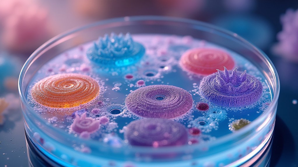
The 7 best specimens for phase contrast imaging include live cell cultures, bacteria, thin tissue sections (5-10 micrometers), protozoa, unstained biological fluids, plant cells, and thin crystalline materials. You’ll achieve ideal results by maintaining appropriate thickness and proper preparation techniques for each specimen type. These samples showcase refractive index differences without staining, allowing visualization of natural cellular structures and dynamic processes. The following guide reveals preparation secrets that transform ordinary samples into stunning phase contrast images.
Live Cell Cultures and Their Optimal Preparation

When preparing live cell cultures for phase contrast microscopy, you’ll need to maintain a thickness between 5 to 10 micrometers to achieve perfect results. Thicker specimens create distortion and halos that obscure cellular details during optimal imaging.
Ensure your cultures are well-adhered to their substrate to minimize movement and allow consistent light passage during observation. Choose appropriate culture media that maintains cell viability while enhancing visibility of structures without staining.
Proper adherence to substrate and media selection: dual keys for stable, high-contrast imaging of unstained live cultures.
Don’t neglect environmental conditions – stable temperature and CO2 levels prevent unwanted changes in cell morphology that could compromise image quality.
Finally, select phase contrast objectives specifically designed for live imaging (look for Ph1, Ph2, or Ph3 green inscriptions) with appropriate numerical aperture values.
This combination of careful preparation and proper equipment will deliver the crisp contrast you need.
Bacterial Specimens and Microbial Communities
Bacterial specimens represent ideal subjects for phase contrast microscopy due to their transparent nature and minimal thickness. You’ll observe live cells in their natural state without staining, allowing you to study motility patterns and behaviors that would otherwise be altered by fixation techniques.
When examining microbial communities like biofilms, phase contrast microscopy reveals complex interactions between different species based on their distinct refractive index properties. This technique differentiates bacterial cells within mixed cultures by highlighting variations in cell morphology and density.
You can identify subcellular structures and inclusions that indicate specific metabolic states, providing deeper insights into bacterial function.
Additionally, phase contrast proves invaluable when researching antibiotic resistance mechanisms, as you’ll clearly visualize morphological changes in response to treatment without the interference of staining artifacts.
Tissue Sections at Ideal 5-10 Micrometer Thickness

Achieving ideal contrast in tissue specimens requires precise sectioning between 5-10 micrometers thick, where cellular structures become remarkably visible without specialized stains.
When you examine tissue slices at this prime thickness under phase contrast microscopy, you’ll notice how the refractive index differences between cellular components create natural contrast.
- Thicker sections (>10 micrometers) produce excessive phase shift, distorting cellular details and compromising image quality.
- Maintaining uniform thickness guarantees consistent contrast across your entire field of view.
- This thickness range preserves specimen integrity, making it perfect for live cell imaging.
- The 5-10 micrometer range provides the perfect balance between visibility and light transmission.
This precise sectioning technique gives you detailed images without chemical stains, making light microscopy more valuable for examining delicate tissue structures.
Protozoa and Single-Celled Organisms
Protozoa and single-celled organisms represent perhaps the most spectacular specimens for phase contrast microscopy.
You’ll find these transparent microbes nearly invisible under standard brightfield techniques, but they transform into detailed, high-contrast subjects when viewed with transmitted light in phase contrast.
Species like *Paramecium* and *Amoeba* become remarkable study subjects without requiring stains that might compromise their viability.
Phase contrast microscopy reveals living protists in stunning detail, preserving natural behaviors unaltered by potentially harmful stains.
The phase contrast technique reveals intricate organelles such as cilia and contractile vacuoles that would otherwise remain hidden.
Your live observations will capture dynamic cellular functions including feeding behaviors and movement patterns in real-time.
This visibility has revolutionized how researchers study microorganisms’ biology and ecological relationships.
Whether you’re teaching microbiology or conducting research, you’ll appreciate how phase contrast brings these otherwise transparent organisms to life with exceptional detail and contrast.
Unstained Biological Fluids and Secretions

You’ll find unstained biological fluids such as urine, saliva, and mucus ideal for phase contrast microscopy since they naturally showcase cellular components without requiring stains.
These specimens benefit from the technique’s ability to transform subtle refractive index differences into visible contrast, revealing leukocytes, crystals, and microorganisms that would otherwise remain hidden.
When examining these fluids under phase contrast, you can observe living cells in their dynamic state, providing valuable insights into cellular behavior and pathological conditions in real-time.
Natural Cellular Visibility
While traditional microscopy often requires staining to reveal cellular details, phase contrast microscopy excels at visualizing unstained biological specimens with remarkable clarity.
You’ll observe cellular components in their native state without the distortion that staining chemicals introduce.
This technique leverages subtle refractive index differences to create stunning visibility in:
- Biological fluids like saliva and urine, where leukocytes and epithelial cells appear in sharp relief
- Live cells suspended in fluid, allowing you to monitor cell motility and interactions in real-time
- Low-contrast specimens including organelles within macrophages, with enhanced detail of their structure
- Microorganisms in liquid media, which become readily distinguishable against clear backgrounds
Phase contrast microscopy truly shines when examining dynamic cellular processes, revealing the natural beauty and complexity of unstained living specimens.
Refractive Index Differences
The magic of phase contrast microscopy lies in its ability to transform invisible differences in optical density into striking visual contrasts.
When you examine unstained biological fluids like saliva, urine, or cell culture media, their natural refractive index differences become readily visible without chemical staining.
Your phase contrast microscope configuration exploits these optical densities to reveal cellular structures and motility in living cells that would otherwise be nearly invisible.
Secretions such as mucus and synovial fluid display distinctive phase variations that highlight their composition and cellular components.
Even blood plasma and lymph reveal their microscopic details through these inherent optical properties.
Living Cell Dynamics
Living cells reveal their intricate dynamics when observed through phase contrast microscopy, offering an unparalleled window into cellular behavior without disrupting natural processes. You’ll achieve high contrast visualization of unstained biological fluids like plasma and cytosol, making real-time analysis of physiological responses possible.
- Watch cell-cell interactions unfold before your eyes, revealing essential mechanisms in immune responses and cancer metastasis.
- Track subtle changes in cellular dynamics during experimental treatments, capturing immediate responses to stimuli.
- Observe secretions like mucus and hormones that would remain invisible in brightfield microscopy.
- Monitor living cells as they divide, move, and change morphology without the artifacts introduced by stains.
This technique transforms nearly invisible phase shifts into striking contrast differences, preserving specimens in their natural state while revealing their functional secrets.
Plant Cells and Transparent Botanical Structures

Because plant cells contain numerous transparent structures, they represent ideal specimens for phase contrast microscopy. You’ll find that without requiring stains, you can clearly visualize cell walls, chloroplasts, and vacuoles that would otherwise remain invisible using conventional techniques.
When working with transparent specimens like leaf epidermis or root hairs, you’re able to observe them in their natural state. This makes phase contrast particularly valuable for studying live plant cells and dynamic cellular processes such as cell division and cytoplasmic streaming. Aim for thin sections (5-10 micrometers) to achieve best contrast without distortion.
The technique’s ability to detect subtle differences in refractive index between cellular components provides valuable insights into plant cell health and physiological status, making it an essential tool for botanical research.
Thin Crystalline Materials and Non-Biological Samples
Moving beyond biological specimens, phase contrast microscopy excels when applied to thin crystalline materials and non-biological samples.
This technique reveals structural details in transparent polymers and crystalline layers without staining, preserving their natural properties while highlighting variations in thickness and density.
You’ll find phase contrast imaging particularly valuable for material science applications when you need to:
- Analyze transparent polymers and thin films to detect subtle defects that remain invisible with conventional microscopy
- Examine the distribution and alignment of crystalline grains to understand mechanical properties
- Study semiconductor materials by visualizing refractive indices differences without sample modification
- Generate high-contrast images that reveal intricate patterns and textures to aid in material characterization
Frequently Asked Questions
What Type of Specimen Is Best for Phase Contrast Microscopy?
You’ll get the best results with phase contrast microscopy when examining thin, live, and unstained specimens like living cells, protozoa, or transparent materials with subtle refractive index differences (5-10 micrometers thick).
How Do You Get a Good Image Under Phase Contrast Microscope?
To get a good phase contrast image, you’ll need a thin specimen (5-10µm), matched Ph objectives and condenser, proper annulus centering, correct aperture settings, and a green filter for enhanced visibility.
What Organism Can Be Seen in a Phase Contrast Microscope?
You can see various organisms like human cheek cells, bacteria (E. coli, B. subtilis), protozoa (Paramecium, Amoeba), and fungi under a phase contrast microscope. It’s excellent for observing live, unstained specimens in their natural state.
What Are the Best Practical Applications of a Phase Contrast Microscope?
You’ll find phase contrast microscopes excel in live cell observation, microbiology research, tissue analysis, forensic investigations, and material science. They’re invaluable when you’re studying transparent specimens without needing to stain or fix them.
In Summary
You’ll achieve exceptional phase contrast imaging by properly preparing these seven specimen types. Whether you’re working with living cells, bacterial communities, or thin tissue sections, remember that sample preparation is critical to your success. Don’t overlook the importance of proper thickness and mounting techniques. With these ideal specimens and careful handling, you’re well-positioned to capture stunning phase contrast images in your microscopy work.


Leave a Reply