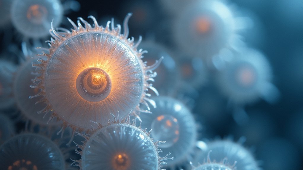
Phase contrast photos require different light values because specimen thickness, density, and transparency affect how light waves diffract. You’ll need higher intensity for transparent specimens and lower for dense ones to prevent overexposure. Proper light calibration prevents haloing effects and reveals cellular structures clearly without stains. Even nanometer misalignments between the condenser’s annular ring and phase plate can compromise image quality. Mastering these adjustments transforms ordinary observations into revealing cellular portraits.
The Optical Principles Behind Phase Contrast Imaging

While invisible to conventional microscopy, transparent specimens come alive through phase contrast imaging. This technique transforms imperceptible phase shifts in light waves into visible amplitude differences, revealing cellular structures without staining.
At its core, phase contrast microscopy employs a specialized optical pathway. You’ll notice the annular diaphragm in your condenser creates a hollow cone of light that passes through your specimen. As these light waves encounter different refractive indices within the sample, they become diffracted.
Your phase contrast objectives contain precisely engineered phase plates that amplify these subtle differences. For successful imaging, you must align your condenser’s annular ring with the phase plate in your objective.
This critical relationship between diffracted light and the direct light beam creates the contrast you need to visualize otherwise transparent features.
How Light Intensity Affects Phase Shift Visualization
When working with phase contrast microscopy, light intensity serves as the foundation for successful visualization of transparent specimens. As you adjust the illumination on your phase contrast microscope, you’re directly influencing the amplitude of light waves interacting with your sample.
Higher light intensity makes phase shifts more pronounced, revealing critical cellular structures that would otherwise remain invisible. You’ll notice considerably improved clarity and definition in your specimens.
Conversely, if your light settings are too low, weak phase contrast will obscure important details and potentially lead to misinterpretation.
For ideal contrast enhancement, you’ll need to carefully calibrate your light source. Whether you’re using halogen or LED illumination, finding the perfect intensity balance prevents both insufficient visualization and excessive brightness that causes glare or saturation in your images.
Optimizing Exposure Settings for Different Specimen Types

When working with transparent specimens, you’ll need to reduce light intensity to prevent excessive scatter, while dense specimens require increased illumination to reveal internal structures.
You can compensate for the haloing effect commonly seen in phase contrast by fine-tuning your condenser’s aperture according to the specimen’s refractive properties.
Remember that proper exposure settings aren’t one-size-fits-all—they must be calibrated to each specimen type to achieve ideal contrast without introducing artifacts.
Transparent vs. Dense Specimens
Because transparent and dense specimens create fundamentally different phase shifts, they require distinct exposure approaches for ideal visualization.
When working with transparent specimens in phase contrast, you’ll need to increase light intensity considerably as these samples (typically 5-10 micrometers thick) have lower inherent contrast and may appear underexposed with standard settings.
Conversely, dense specimens scatter light more effectively due to their higher refractive index differences. You’ll want to reduce your light values when examining these samples to avoid overexposure that obscures essential details.
Remember that finding the right balance is vital—too much light on dense specimens creates saturation while insufficient illumination leaves transparent structures invisible.
Haloing Effect Compensation
The characteristic bright rings surrounding darker specimen outlines—known as the haloing effect—represent one of phase contrast microscopy’s most significant challenges.
To compensate for this optical artifact, you’ll need to carefully adjust your exposure settings based on specimen thickness and transparency.
For thinner specimens, increasing light values can help overcome haloing while preserving detail. Conversely, thicker samples require reduced intensity to prevent overexposure in high phase shift areas. Living cells demand particularly precise calibration compared to fixed tissues due to their variable refractive properties.
Using objectives with higher numerical aperture improves resolution but necessitates further fine-tuning of light values to avoid saturation.
Regular alignment of your phase contrast setup—ensuring proper matching between condenser annulus and phase rings—remains essential for consistent results that effectively minimize haloing while maintaining ideal contrast.
Balancing Contrast and Resolution in Phase Microscopy
When working with phase contrast microscopy, you’ll find that adjusting the illumination angle considerably affects how light interacts with your specimen, often determining whether subtle structures remain visible or disappear.
You can manipulate the balance between amplitude (brightness) and phase (optical path differences) by fine-tuning your light source intensity and condenser position.
These adjustments will help you achieve the ideal compromise between high contrast and maximum resolution, particularly when imaging transparent specimens with minimal inherent contrast.
Illumination Angle Effects
Finding the perfect illumination angle stands as one of the most critical factors in phase contrast microscopy, where even slight adjustments can dramatically alter image quality.
When you’re working with phase contrast, you’ll notice that higher illumination angles may enhance edge details but often introduce unwanted halos around areas with significant phase shifts.
As you adjust your microscope’s light values, you’ll need to take into account the specimen thickness. Thicker specimens particularly suffer from distortion under improper illumination conditions, causing resolution loss.
You’ll achieve ideal results by carefully aligning your phase contrast condenser with your objectives, preventing mismatched illumination angles that degrade image quality.
Regular calibration of your illumination system isn’t just recommended—it’s essential for maintaining that delicate balance between contrast and resolution that defines excellent phase contrast microscopy.
Amplitude vs. Phase
Understanding the fundamental interplay between amplitude and phase represents the cornerstone of effective phase contrast microscopy. When you’re using this contrast technique, you’re fundamentally converting phase shifts (caused by refractive index differences) into amplitude variations that your eye can detect.
In light microscopy, amplitude refers to light intensity while phase denotes wave position. You’ll need to balance both elements carefully—improper settings often create distracting halos that obscure fine cellular details.
Your microscope’s numerical aperture alignment between the phase ring and condenser annulus directly affects how effectively phase differences translate into visible contrast.
Select appropriate light wavelengths for your specimen’s refractive properties to enhance structural visibility. Without this balance, you’ll sacrifice either contrast or resolution, limiting your ability to clearly visualize unstained biological samples without introducing artifacts.
Common Light Value Adjustments for Cellular Imaging

To achieve ideal results in phase contrast microscopy, proper light value adjustment stands as the cornerstone of clear cellular imaging.
You’ll need to calibrate light intensity carefully—too much brightness enhances contrast but introduces unwanted halos around high phase shift areas of your specimen.
Always match your condenser’s numerical aperture with your objective lens and light source intensity. This alignment is essential for best phase contrast imaging.
Consider using monochromatic light sources when possible, as consistent wavelengths maintain more reliable phase relationships and improve image clarity.
Different specimen types and thicknesses require specific light adjustments.
You should regularly realign your microscope’s optical components to adapt to these variables, ensuring your phase contrast photos consistently reveal cellular structures without requiring stains or dyes.
Troubleshooting Image Quality Issues Through Light Calibration
Most image quality problems in phase contrast microscopy stem from improper light calibration. When you notice halo artifacts obscuring cellular details, you’ll need to adjust your light intensity to reduce these optical distortions while maintaining sufficient contrast to visualize structures.
Check that your condenser’s numerical aperture matches your objective—mismatched values considerably degrade image quality.
Verify your phase rings are properly aligned; even slight misalignments can dramatically reduce high contrast in your specimens.
If you’re still experiencing issues, recalibrate your light settings based on your specific specimen’s thickness and type. Different cells require different light values to optimize visualization.
Remember that your light source matters too—halogen or LED illumination should be properly aligned to provide even illumination across the entire field, enhancing overall phase contrast quality.
Advanced Techniques for Enhancing Phase Contrast Detail

While basic phase contrast delivers adequate images, mastering advanced light value manipulation will dramatically improve your visualization of cellular structures.
When working with thick specimens, you’ll need to adjust light intensity to penetrate effectively without washing out subtle details. Try pairing your light microscope settings with the specific optical properties of your sample to minimize halo artifacts at high-contrast boundaries.
Ensure perfect alignment between your condenser annulus and objective’s numerical aperture—even slight mismatches create significant differences in image quality.
Precise condenser-objective alignment is the cornerstone of exceptional microscopy—even nanometer deviations can dramatically alter visualization results.
You can fine-tune wavelength selection to enhance specific cellular features; shorter wavelengths often reveal finer details in transparent specimens.
For dynamic processes, optimize light values in real-time, balancing contrast against artifact formation. This calibration allows you to observe living cells without staining, preserving their natural behavior during observation.
Frequently Asked Questions
What Happens to Light Rays in a Phase Contrast Microscope?
In a phase contrast microscope, light rays pass through your specimen, undergo phase shifts due to refractive index differences, and are converted into amplitude differences by the phase plate, creating visible contrast.
What Light Does Phase Contrast Microscope Use?
You’ll find that phase contrast microscopes typically use halogen or LED light sources. They’re designed to provide bright, stable illumination that’s essential for highlighting phase differences in transparent specimens without staining.
How to Obtain a Good Image Under a Phase Contrast Microscope?
You’ll need to align the phase rings precisely, adjust light intensity for ideal contrast, use appropriate objective lenses, and guarantee your specimen thickness is 5-10 micrometers for clear phase contrast images.
Why Is Green Light Used in Phase Contrast Microscopy?
You’ll find green light (500-550nm) ideal in phase microscopy because it matches your eye’s sensitivity, reduces aberrations, enhances contrast in transparent specimens, and minimizes halos around high phase shift areas.
In Summary
You’ve now mastered the fundamentals of phase contrast photography and understand why proper light values are essential. By adjusting your illumination settings for different specimens, you’ll capture the subtle phase shifts that reveal otherwise invisible structures. Remember, there’s no one-size-fits-all approach—you’ll need to experiment with your light values to find the perfect balance between contrast and resolution for each unique subject you photograph.


Leave a Reply