
Positive phase contrast microscopy makes your cells appear bright against a dark background, enhancing visibility of organelles and membrane boundaries without staining. You’ll see nuclei, mitochondria, and other structures clearly while cells remain alive and undisturbed. It’s non-invasive, preserving cellular integrity while providing exceptional detail of transparent specimens. The technique transforms phase shifts into brightness differences, making it superior for observing cell morphology and dynamic processes. Discover how this technique revolutionizes your live cell imaging capabilities.
The Fundamentals of Positive Phase Contrast Microscopy
Visibility is the cornerstone of cellular observation, and positive phase contrast microscopy revolutionizes how we see living cells. This contrast technique enhances cellular structures by advancing light waves that pass through the specimen, creating bright images against dark backgrounds.
When you’re examining unstained specimens, positive phase contrast capitalizes on differences in refractive indices between cell components and their surroundings. The system transforms subtle optical path variations into dramatic brightness differences, making cell nuclei and organelles clearly visible without staining.
The setup requires specialized objectives and a condenser with an annular aperture that shapes light precisely. This configuration allows you to observe live cells in real-time while maintaining their integrity—a critical advantage for studying dynamic cellular processes in microbiology and cytology research.
Enhanced Visualization of Cellular Structures and Boundaries
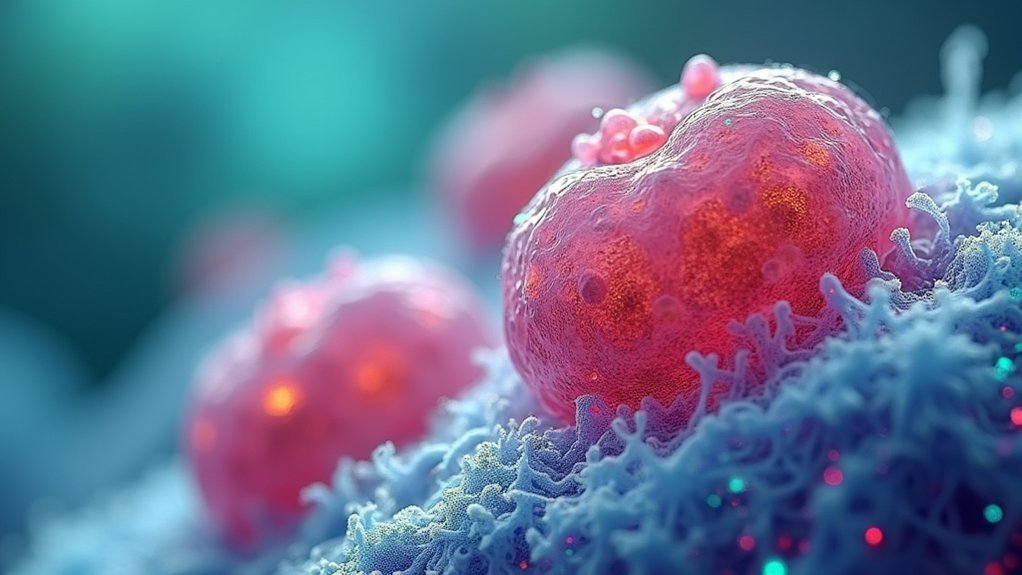
When examining living cells under positive phase contrast microscopy, you’ll immediately notice how cellular structures transform into luminous features against a dark backdrop. This contrast technique enhances visualization by advancing light waves from specimens, making cell boundaries remarkably distinct and easier to observe.
You’ll appreciate how positive phase contrast reveals organelles and intracellular components that might remain hidden with other microscopy methods. The nucleus, mitochondria, and other essential structures become clearly discernible without requiring chemical stains that could compromise cell health or function.
This advanced approach is particularly valuable when you’re studying unstained living cells in real time. By providing superior differentiation between cellular elements and their surroundings, you’ll gain more accurate insights into cell morphology, behavior, and dynamic processes such as division and motility.
Live Cell Imaging Without Compromising Cellular Integrity
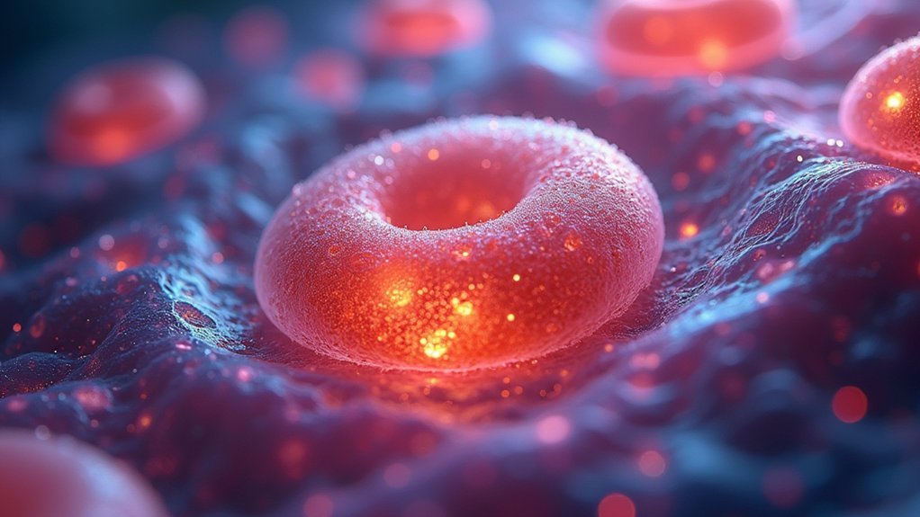
Positive phase contrast microscopy offers a remarkable advantage for researchers studying live cells: you can observe cellular processes without disrupting the specimen’s natural state.
This non-invasive technique converts phase shifts into visible contrast differences, allowing you to examine delicate organelles and cellular structures in their natural environment.
Key benefits of this approach include:
- Preservation of cellular integrity during extended observation periods, enabling real-time monitoring of dynamic processes
- High-contrast imaging of thin specimens (5-10 micrometers) without introducing staining artifacts
- Enhanced visibility of live cells against dark backgrounds, making it invaluable for microbiology applications
You’ll appreciate how positive phase contrast maintains cellular integrity while delivering exceptional detail—a critical factor when studying functional, living systems rather than fixed specimens.
Comparing Positive vs. Negative Phase Contrast for Cell Observation
Although both methods reveal cellular details without staining, positive and negative phase contrast techniques differ greatly in how they present cellular structures to observers.
When you use positive phase contrast, transparent specimens appear brightly illuminated against a darker background, making cell structures more pronounced. The bright halos created enhance visibility of organelles and fine details within cells.
Negative phase contrast reverses this relationship, showing darker cells against a lighter field.
You’ll find that positive phase contrast provides superior contrast enhancement for observing cell morphology and dynamic cellular processes in real-time. This imaging technique is particularly valuable when studying living biological specimens, as you can visualize cellular interactions and behaviors with greater clarity than with negative phase contrast, making it the preferred choice for many researchers.
Optimizing Microscope Settings for Cellular Phase Contrast Imaging
Successful phase contrast imaging of cells depends on precise microscope configuration that maximizes structural visibility while minimizing artifacts.
To obtain high-quality phase contrast images, you’ll need to properly align the annular diaphragm with the objective’s phase ring, ensuring ideal contrast for visualizing cell structures.
Precise alignment of the annular diaphragm with the phase ring creates optimal contrast, revealing cellular details invisible to standard microscopy.
For ideal results:
- Set up Köhler illumination to create uniform specimen lighting, greatly reducing unwanted artifacts while enhancing contrast.
- Choose appropriate phase contrast objectives (look for Ph1, Ph2, Ph3 markings) that match your specimen’s thickness and transparency requirements.
- Adjust your light source intensity carefully—too bright washes out details, while too dim obscures fine cellular features.
Regular cleaning of optical components prevents contamination that could degrade image quality and compromise the phase contrast effect.
Applications of Positive Phase Contrast in Modern Cell Biology Research
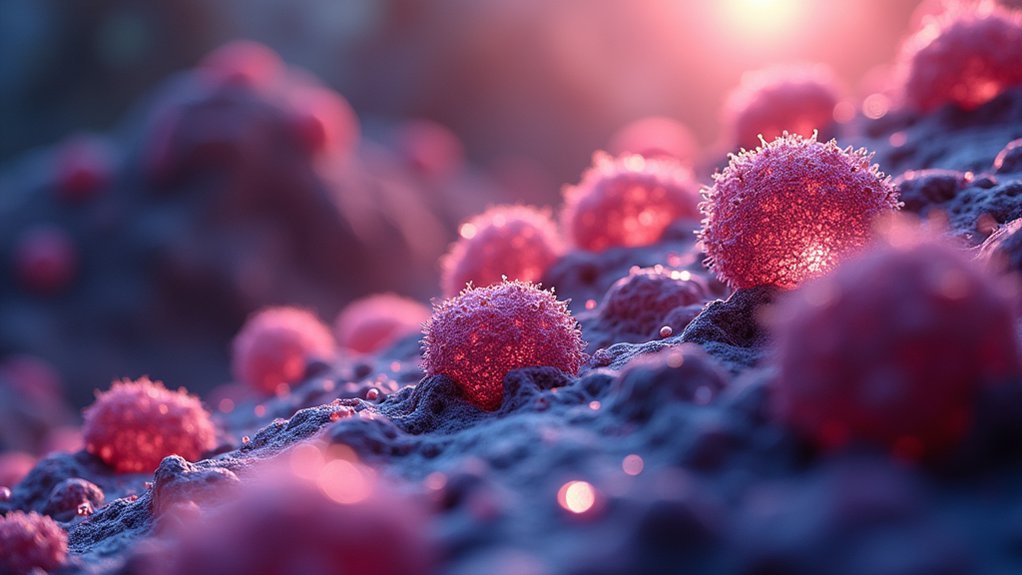
In contemporary cell biology research, positive phase contrast microscopy has revolutionized how scientists visualize and study living cells without disrupting their natural functions.
You’ll find this technique indispensable in cytology when evaluating cell morphology and viability for cancer diagnostics.
In microbiology, positive phase contrast reveals bacterial behaviors and structures that would remain invisible with conventional microscopy.
When studying transparent specimens, you’ll observe dynamic processes like cell division and differentiation in real time—something impossible with fixed, stained samples.
Researchers working on drug development rely on this non-invasive approach to monitor cellular interactions and responses to treatments.
Frequently Asked Questions
What Is the Difference Between Positive and Negative Phase Contrast?
Positive phase contrast shows cells as bright against a dark background by advancing light phases, while negative phase contrast displays them as dark against a bright background by retarding light phases. You’ll see different contrasts with each method.
Why Is Phase Contrast Better Than Bright-Field?
Phase contrast outperforms bright-field because you can visualize transparent, unstained living cells clearly. It converts phase shifts into visible contrast differences, revealing cellular structures and dynamic processes that you’d completely miss with standard bright-field illumination.
What Are the Advantages of Phase Contrast?
Phase contrast gives you clear views of unstained living cells, revealing structures invisible in bright-field. You’ll detect subtle details through refractive index differences, while avoiding staining artifacts that might damage your specimens.
How Is Negative Phase Different From Positive Phase?
In negative phase contrast, you’ll see cells appear dark against a bright background, while in positive phase, they’re bright against a dark background. This difference affects how clearly you can view cellular structures.
In Summary
When you’re working with living cells, positive phase contrast is your best ally. You’ll see crisp boundaries and internal structures as bright images against a darker background, preserving cellular integrity without staining. It’s superior to negative contrast for most biological applications, offering better definition and natural appearance. Just adjust your microscope properly, and you’ll reveal detailed, real-time observations essential for modern cell biology research.


Leave a Reply