Digital phase contrast image analysis combines traditional phase contrast microscopy with advanced digital technologies to visualize and quantify living cells without chemical staining. You’ll see remarkable advancements in this field through deep learning algorithms that achieve correlation coefficients up to 0.953 in cell quantification. Modern systems feature real-time processing capabilities, automated cell tracking, and sophisticated artifact correction. The integration of high-resolution cameras with specialized software transforms cellular research with unprecedented accuracy and efficiency. The evolution continues with each technological breakthrough.
What Is Digital Phase Contrast Image Analysis Today?

While traditional imaging techniques often require cell staining that can disrupt biological processes, digital phase contrast image analysis stands out as a non-invasive method that revolutionizes how researchers visualize and quantify living cells.
Digital phase contrast analysis reveals living cells without disrupting their natural state—illuminating biology’s true dynamics.
You’ll find this technology leverages sophisticated algorithms and convolutional neural networks (CNNs) to transform raw microscopy data into actionable insights.
When you’re conducting cell counting, this technique offers remarkable precision without the interference of chemical labels. The technology continuously improves as researchers feed more training images into the system—studies show significant reductions in mean squared error for cell lines like A549 and Huh7 with increased training data.
Advanced restoration algorithms also help overcome common artifacts like halo and shade-off effects that previously limited accurate segmentation, enabling real-time observation of dynamic cellular processes.
The Evolution of Phase Contrast Microscopy in the Digital Era
Phase contrast microscopy has undergone a remarkable transformation since its inception in the early 20th century.
Today, you’ll find digital imaging technology seamlessly integrated into these systems, dramatically enhancing resolution and clarity when examining living cells.
The digital revolution has introduced powerful computational algorithms that automate image analysis, making cell counting and tracking more accurate than ever before.
You can now observe dynamic cellular processes in real-time without staining your specimens, opening new possibilities in developmental biology research.
High-throughput capabilities have become a cornerstone of modern phase contrast microscopy, with high-content screening enabling simultaneous analysis of multiple samples.
Your research workflow benefits from advanced digital cameras and specialized software that quantify subtle morphological changes, transforming how you visualize and interpret cellular behavior.
Fundamental Principles of Digital Phase Contrast Analysis

At the core of digital phase contrast analysis lies the ingenious transformation of invisible phase shifts into detectable amplitude differences. When you use a phase contrast microscope, you’re leveraging Zernike’s optical principles—specifically annular apertures and phase plates that manipulate light waves to reveal transparent structures without staining.
The digital evolution enhances these capabilities by applying sophisticated algorithms that convert subtle phase information into clear visual data. Modern segmentation methods overcome traditional challenges like halo artifacts and shade-off effects through restoration algorithms.
What’s particularly exciting is how machine learning, especially CNNs, now automates cell counting with improved accuracy, reducing MSE across various cell lines.
This digital approach integrates seamlessly with High Content Screening, enabling you to monitor living cells in real-time while maintaining sample integrity—a critical advantage for dynamic biological studies.
Hardware Components for Advanced Phase Contrast Imaging
The backbone of any high-performance phase contrast imaging system consists of several precisely engineered components working in harmony.
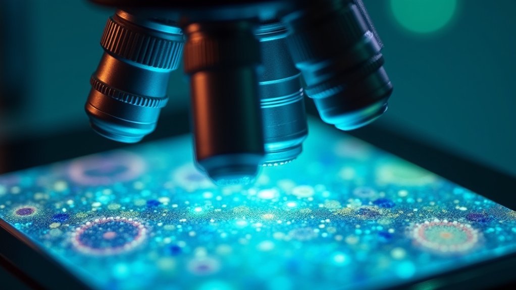
When you’re setting up your imaging station, you’ll need specialized phase contrast microscopes with objectives designed to enhance contrast in transparent specimens through light phase shifts.
For the best results, verify your system includes:
- High-quality halogen or LED light sources that deliver consistent illumination while minimizing photobleaching in your living specimens
- Precisely calibrated condensers with annular apertures that properly modify the light path
- High-resolution digital cameras capable of capturing images for quantitative analysis
- Calibration tools and software to maintain perfect alignment of optical components
These hardware components work together to transform invisible phase differences into visible contrast, allowing you to explore cellular structures with unprecedented clarity.
Computational Algorithms Enhancing Phase Contrast Visualization

While hardware components provide the foundation for phase contrast imaging, sophisticated computational algorithms have transformed how we visualize and interpret these images.
You’ll find that regularized quadratic optimization techniques now restore artifact-free images, effectively addressing common issues like halo and shade-off effects that previously hindered analysis.
These computational algorithms employ linear imaging models that accurately represent the optical properties of phase contrast microscopy systems.
Linear imaging models within computational algorithms precisely mirror phase contrast microscopy’s optical characteristics
By integrating machine learning with advanced image processing, you can analyze dynamic cellular processes in real-time without staining—preserving sample integrity while improving detection accuracy.
Enhanced segmentation methods incorporating these restoration algorithms notably improve quantification of cell populations.
This integration has revolutionized biological research by providing cleaner data for neural network models, ultimately enabling more precise tracking and analysis of living specimens.
Deep Learning Applications in Phase Contrast Image Interpretation
You’ll find CNN architecture optimization critical for accurate cell quantification, as evidenced by A549 cell line models achieving an impressive MSE of 4,335.99 and correlation coefficient of 0.953.
Automated feature extraction eliminates the need for cell staining while maintaining high accuracy across varying cell densities, though 3T3 cell lines still present challenges due to poor signal-to-noise ratios.
Multi-scale pattern recognition, enhanced through data augmentation techniques like rotation and scaling, has transformed limited datasets into robust training resources, considerably improving deep learning model performance in phase contrast image interpretation.
Neural Network Architecture Optimization
Designing ideal neural network architectures represents a critical step in maximizing the performance of deep learning systems for phase contrast image analysis.
You’ll find CNN architectures particularly effective for cell counting applications, with A549 cell models achieving impressive MSE values of 4,335.99 compared to other cell lines.
By implementing data augmentation techniques like rotation and scaling, you can dramatically increase your training dataset robustness.
- Watching your model achieve near-perfect correlation coefficients (R=0.953) when architecture optimization succeeds
- Feeling frustrated when image artifacts disrupt even well-designed neural networks
- Experiencing breakthrough moments when optimization algorithms overcome persistent convergence issues
- Celebrating when your optimized architecture finally handles challenging halo effects in phase contrast images
Automated Feature Extraction
Automated feature extraction represents the practical application of optimized neural networks in phase contrast microscopy. You’ll find CNNs at the forefront of this technology, excelling at identifying spatial patterns in cellular structures without staining requirements.
These networks process phase contrast light information to perform accurate cell counting with impressive precision, as evidenced by MSE values of 4,335.99 for A549 cell lines.
Your results will vary by cell type—A549 and Huh7 lines demonstrate strong correlations (R = 0.953 and R = 0.821) while 3T3 cells present challenges (R = 0.100).
To improve model robustness, data augmentation techniques have expanded training datasets substantially, transforming 795 A549 images into 2,673. Advanced restoration algorithms now target halo and shade-off artifacts, enhancing segmentation accuracy and enabling reliable cell detection in automated systems.
Multi-Scale Pattern Recognition
Deep learning approaches have revolutionized multi-scale pattern recognition in phase contrast imagery, particularly through their ability to detect features across varying cellular dimensions.
You’ll find CNNs at the forefront, progressively learning to identify complex structures while achieving impressively low MSE values—exemplified by the A549 cell line’s 4,335.99 measurement.
Data augmentation techniques transform modest datasets into robust training collections, as demonstrated when the A549 database expanded from 167 to 2,673 images through rotation and scaling adjustments.
- Witness correlation coefficients reaching 0.953 between predicted and actual cell counts!
- Marvel at how machines can now interpret what once required expert human analysis
- Experience frustration with persistent challenges in analyzing dense cell populations
- Feel optimistic about future breakthroughs in low signal-to-noise scenarios
Quantitative Analysis Methods for Cell Morphology and Dynamics
As researchers investigate deeper into cellular behavior, quantitative analysis of cell morphology and dynamics has become essential for extracting meaningful data from digital phase contrast images.
You’ll find advanced algorithms that measure critical parameters including cell area, perimeter, and circularity—providing objective insights into cellular characteristics.
Machine learning techniques, particularly CNNs, have revolutionized this field by greatly improving accuracy in cell counting and morphological assessment.
With error values now as low as 4,335.99 in specific cell lines, you can track cell shape changes and movement over time with unprecedented precision.
High Content Screening has further transformed large-scale studies by rapidly evaluating thousands of images.
Real-Time Processing Techniques for Live Cell Monitoring

While traditional microscopy methods require fixing and staining cells, real-time processing in digital phase contrast imaging now allows you to monitor living cells continuously without compromising their viability.
Advanced algorithms powered by machine learning enhance image quality instantly, correcting halo artifacts and improving contrast as you watch. You’ll capture rapid cellular events like division and movement as they unfold, enabling immediate analysis of dynamic processes.
- Witness the miracle of cell division happening before your eyes with high-speed imaging capabilities
- Feel the excitement of discovering cellular interactions the moment they occur
- Experience the satisfaction of seeing crystal-clear images without damaging your precious samples
- Transform your research with immediate feedback rather than waiting hours for results
Comparison Between Traditional and Digital Phase Contrast Systems
Traditional phase contrast microscopes have evolved from purely optical systems into sophisticated digital platforms with advanced algorithms that eliminate artifacts and enhance cellular details.
You’ll notice dramatic improvements in performance metrics when comparing digital systems to their traditional counterparts, particularly in resolution quality, processing speed, and the accuracy of quantitative measurements.
Modern digital phase contrast technology delivers superior image analysis capabilities through machine learning integration, enabling automated cell tracking and real-time monitoring that weren’t possible with conventional systems.
Equipment Evolution Comparison
Over the past decade, phase contrast microscopy has undergone a remarkable transformation from purely optical systems to sophisticated digital platforms. Where you once needed specialized annular diaphragms and phase plates, you’ll now find powerful image processing algorithms driving digital phase contrast systems that quantify cellular structures with unprecedented precision.
- Breakthrough speed – analyze thousands of images in minutes instead of hours
- Freedom from frustration – automatic correction of halo and shade-off artifacts
- Democratized analysis – user-friendly interfaces making complex technology accessible
- AI-powered insights – machine learning algorithms that count and classify cells with superhuman accuracy
This evolution has democratized advanced microscopy, allowing you to achieve reliable, high-quality results regardless of your technical expertise, while considerably reducing the time spent on manual analysis tasks.
Performance Metrics Analysis
As researchers meticulously compare traditional and digital phase contrast systems, quantitative performance metrics reveal a striking advantage for digital approaches. You’ll notice significant improvements in cellular analysis when using advanced digital methods that minimize artifacts while maximizing resolution.
| Performance Metric | Traditional Systems | Digital Systems |
|---|---|---|
| Mean Squared Error | Higher values | Lower values |
| Image Segmentation | Manual/challenging | Automated/ML-enhanced |
| Analysis Speed | Time-intensive | Real-time processing |
| Artifact Reduction | Limited capability | Advanced algorithms |
| Sensitivity | Moderate | High precision |
Digital systems excel particularly when processing complex cell images, applying machine learning techniques that traditional microscopy simply cannot match. You’ll benefit from immediate feedback on cell morphology and dynamics, drastically improving your experimental workflow and ability to detect subtle changes in live cell behavior with enhanced sensitivity and specificity.
Clinical and Research Applications of Digital Phase Contrast Analysis

While many imaging technologies require complex sample preparation, digital phase contrast analysis has emerged as a cornerstone methodology in both clinical medicine and scientific research.
You’ll find this technique invaluable for identifying cell regions without stains, allowing real-time observation of cellular dynamics across diverse applications.
- Watch cancer cells transform and migrate before your eyes, revealing critical insights into metastasis
- Witness bacterial communities evolve and interact in real-time, revolutionizing infectious disease research
- See immediate cellular responses to potential therapeutics, accelerating drug discovery timelines
- Unveil quantitative data from living systems that would otherwise remain invisible
The integration with machine learning algorithms has further enhanced the technology’s capability to extract meaningful data from complex biological systems, making it indispensable in modern biomedical research.
Frequently Asked Questions
What Is Digital Phase Contrast?
Digital phase contrast is a microscopy technique that you’ll find enhances transparent specimens’ visibility. It converts phase shifts into viewable images using specialized optics and digital processing for better analysis of biological structures.
What Is a Phase Contrast Image?
A phase contrast image is what you’ll see when transparent specimens are made visible by converting light phase shifts into amplitude differences. You’re able to observe living cells without staining through this microscopy technique.
When Would You Use the Phase Contrast Microscope?
You’d use phase contrast microscopes when you need to observe living, unstained cells. They’re perfect for monitoring bacteria, tracking cell division, evaluating culture health, and examining transparent specimens without disrupting their natural state.
What Is the Phase Contrast Microscope Analysis?
Phase contrast microscope analysis is a technique you’ll use to examine transparent specimens. You’re analyzing the contrast differences created when light passes through cells, allowing you to see unstained structures with enhanced visibility.
In Summary
You’ve witnessed the remarkable transformation of phase contrast imaging through digital integration. You’re now equipped with tools that offer unprecedented quantitative analysis and real-time visualization capabilities. As you continue your research or clinical work, you’ll find these advanced systems dramatically enhance your ability to study cellular dynamics with precision that was unimaginable in traditional microscopy. The future of digital phase contrast analysis is already in your hands.


Leave a Reply