The best phase contrast objectives include Zeiss Plan-Neofluar Ph3 100x for high magnification work, Nikon CFI Plan Apochromat Lambda 40X Ph2 for superior resolution, and Olympus UPlanFLN 20X for versatility. Budget-friendly options like Leica HCX PL FLUOTAR 10X/0.30 PH1 deliver quality at lower cost. For specialized needs, consider long working distance objectives for thick specimens. Proper maintenance extends their lifespan considerably. Our detailed breakdown reveals which objectives deliver the most value for different research applications.
Zeiss Plan-Neofluar Ph3 100x Oil Immersion Objective
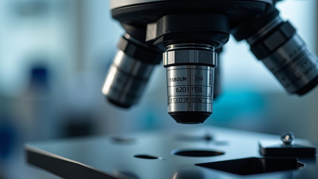
When examining cellular structures at the highest magnification, you’ll find the Zeiss Plan-Neofluar Ph3 100x Oil Immersion Objective delivers exceptional performance for serious microscopists.
This premium phase objective provides remarkable contrast for transparent specimens without staining, revealing intricate details invisible with conventional microscopy.
With its impressive 1.3 numerical aperture, this objective maximizes light collection and resolution capabilities of your phase contrast microscope.
The oil immersion design considerably reduces spherical aberrations that typically limit image quality at high magnifications.
Enhanced optical precision with minimal distortion—where demanding science meets crystal-clear cellular visualization.
You’ll appreciate the Plan-Neofluar’s flat field technology, which eliminates distortion across your entire viewing area—critical for accurate measurements and detailed observation.
It seamlessly integrates with standard phase rings and condensers, making it a worthwhile investment for researchers demanding precision and clarity in high-resolution cellular imaging.
Nikon CFI Plan Apochromat Lambda 40X Ph2 Objective
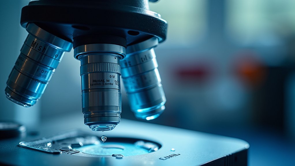
You’ll experience remarkable clarity with Nikon’s CFI Plan Apochromat Lambda 40X Ph2 Objective, which delivers superior optical performance through its high 1.30 numerical aperture.
The plan apochromat design guarantees perfect flatness field correction, eliminating distortion across your entire viewing area.
Its phase contrast capabilities strike an ideal image contrast balance, revealing transparent specimens without staining—crucial for maintaining cell viability during extended observations.
Superior Optical Performance
Precision engineers at Nikon have crafted the CFI Plan Apochromat Lambda 40X Ph2 Objective to deliver exceptional image clarity that justifies its premium position. You’ll immediately notice the superior optical performance when viewing transparent specimens, with minimized chromatic and spherical aberrations that reveal fine structural details.
| Feature | Benefit | Application |
|---|---|---|
| 1.30 NA | Higher resolution | Fine cellular details |
| Advanced coatings | Reduced reflections | Clearer image quality |
| Lambda series design | Superior color fidelity | Accurate specimen representation |
| Ph2 phase contrast | Enhanced transparency | Unstained biological samples |
The objective’s remarkable light-gathering capability transforms your phase contrast microscopy, making previously invisible structures clearly visible. When you’re examining delicate biological specimens that require precise imaging without staining, this objective delivers the exceptional performance serious researchers demand.
Flatness Field Correction
Beyond optical excellence, the Nikon CFI Plan Apochromat Lambda 40X Ph2 Objective offers exceptional flatness field correction that revolutionizes your microscopy experience.
You’ll immediately notice uniform focus across your entire field of view, eliminating the frustration of constantly readjusting when examining different areas of your specimen.
The Lambda design brings remarkable benefits for visualizing phase objects with precision:
- Edge-to-edge clarity – No more blurry peripheries that compromise data collection from your entire sample
- High contrast imaging – Transparent specimens appear with striking definition throughout the field
- Consistent results – Your research gains reliability with uniformly focused images that capture every critical detail
This objective’s advanced flatness field correction technology guarantees you’re not missing crucial information hiding in poorly focused regions of your specimens.
Image Contrast Balance
The remarkable image contrast balance of Nikon’s CFI Plan Apochromat Lambda 40X Ph2 Objective transforms how you observe transparent specimens.
With its impressive 0.95 numerical aperture, you’ll capture subtle details in unstained samples that would otherwise remain invisible.
What sets this objective apart is its advanced coating technology that minimizes stray light interference while maximizing the phase shift visualization.
You’ll notice exceptional clarity as the objective reduces both spherical and chromatic aberrations, delivering crisp images with true-to-life contrast.
Whether you’re using an inverted or upright microscope, this versatile objective integrates seamlessly with Nikon’s phase contrast systems.
You’ll appreciate the precise alignment and enhanced contrast when examining live cells or other delicate specimens that require non-invasive imaging techniques.
Olympus UPlanFLN 20X Phase Contrast Objective
Scientists seeking exceptional cellular detail will find the Olympus UPlanFLN 20X Phase Contrast Objective a worthwhile investment. With its impressive 0.45 numerical aperture, this objective excels at revealing intricate structures in transparent specimens without requiring stains.
The objective’s advanced optical design creates ideal phase contrast microscopy by precisely manipulating light at the objective rear focal plane.
You’ll appreciate these standout features:
- Versatile compatibility with both upright and inverted microscope setups
- Generous 2.0 mm working distance for thicker samples and in-microscope manipulations
- Premium glass construction with specialized coatings that minimize aberrations
Whether you’re conducting live cell imaging or examining delicate cellular structures, this objective delivers the clarity and resolution you need for accurate observations and analysis.
Leica HCX PL FLUOTAR 10X/0.30 PH1 Objective

For researchers seeking a versatile entry-level phase contrast solution, Leica’s HCX PL FLUOTAR 10X/0.30 PH1 Objective delivers exceptional value.
This objective excels in phase contrast microscopy, allowing you to observe live specimens without staining procedures that might affect cellular viability.
The integrated phase plate creates precise phase shifts that transform transparent biological samples into high-contrast images, revealing intricate cellular structures that would otherwise remain invisible in standard light microscopy.
With its 0.30 numerical aperture, you’ll enjoy enhanced light-gathering capabilities critical for detailed observations.
The 10X magnification strikes an ideal balance between detail and field of view, making this objective particularly useful for examining larger specimens.
The FLUOTAR optical design minimizes aberrations, ensuring you’ll consistently obtain clear, accurate images of your unstained specimens.
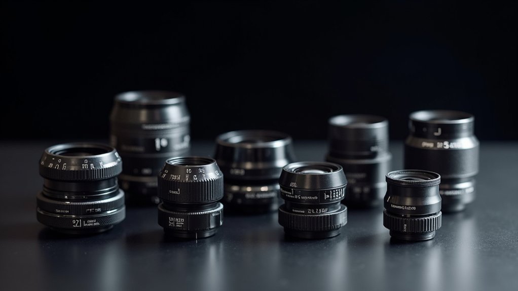
High-Performance Budget Options for Research Microscopy
While acquiring cutting-edge optical equipment often strains laboratory budgets, today’s high-performance budget phase contrast objectives offer remarkable value without compromising essential imaging capabilities.
These affordable options feature quality phase rings that enhance visualization of transparent specimens while maintaining excellent numerical apertures between 0.25 and 0.65.
- Cost-effective objectives with anti-reflective coatings greatly reduce glare, improving image clarity in contrast microscopy applications.
- Compatible components work seamlessly with standard microscope models, making upgrades accessible for labs with limited funding.
- Enhanced resolution allows you to observe live cells and delicate structures with surprising detail, supporting sophisticated research without the premium price tag.
You’ll find these budget-friendly alternatives deliver professional-quality results while preserving your research funds for other critical investments.
Specialized Phase Objectives for Live Cell Imaging
When selecting specialized phase objectives for live cell imaging, you’ll find long working distance options particularly valuable for observing cultures in deep vessels without compromising image quality.
Advanced models featuring chromatic aberration correction deliver sharper images across multiple wavelengths, essential for accurate interpretation of cellular structures and behaviors.
Multi-phase ring designs offer unprecedented versatility by allowing you to switch between different contrast methods without changing objectives, saving time during critical time-lapse experiments.
Long Working Distance Options
Three critical considerations make long working distance phase contrast objectives worth your investment for live cell imaging.
When observing thick specimens, these specialized objectives provide the necessary space between the lens and your cells, preventing damage while maintaining clarity. With NA values between 0.4 and 0.65, they strike the perfect balance between resolution and working distance.
- Enhanced observation capabilities – The integrated phase annulus dramatically improves contrast in live cells without sacrificing image quality.
- Greater specimen flexibility – Ideal for tissue cultures and other thicker samples that traditional objectives struggle to image properly.
- Improved research outcomes – You’ll capture dynamic cellular processes with remarkable clarity, advancing your biological research considerably.
Chromatic Aberration Correction
Despite their higher price point, specialized phase objectives with chromatic aberration correction deliver unparalleled image quality that’s essential for precise live cell imaging.
These objectives maintain a uniform refractive index across the spectrum, ensuring all light wavelengths converge at the same focal point.
When you’re examining transparent samples, the phase ring in these objectives works consistently across different colors, preventing the rainbow-effect distortions that can obscure critical cellular details.
Advanced optical coatings and specialized glass formulations enhance how light phase is maintained throughout the optical path.
You’ll notice the difference immediately in color fidelity and contrast when observing unstained specimens.
This investment translates directly to higher-quality data and more accurate observations of cellular structures and dynamics—crucial advantages for serious research applications.
Multi-Phase Ring Designs
Revolutionary in their design, multi-phase ring objectives represent the cutting edge of live cell imaging technology.
These specialized objectives utilize multiple phase annuli to enhance contrast in unstained specimens, allowing you to observe cellular structures with remarkable clarity. You’ll notice considerably reduced halo effects, making it easier to distinguish fine details during critical observations.
When considering these advanced objectives for your microscopy setup, note their key advantages:
- Enhanced visualization of subcellular components and dynamic processes like cell division
- Versatile compatibility across different magnifications for various specimen types
- Superior contrast without requiring potentially harmful staining procedures
Investing in multi-phase ring designs will dramatically improve your live cell imaging capabilities, providing the clarity and detail necessary for advanced biological research and analysis.
Essential Maintenance Tips to Maximize Objective Lifespan
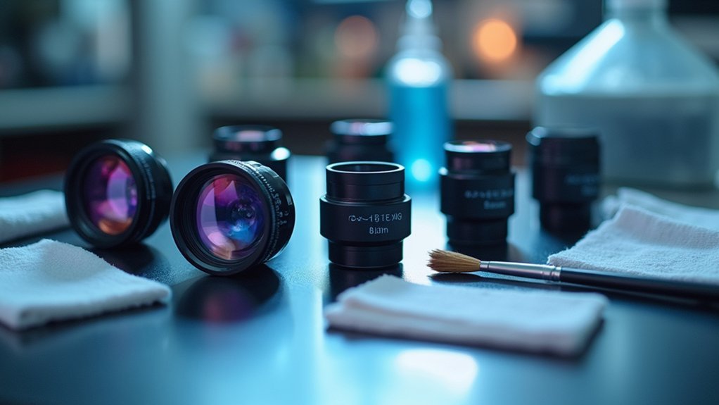
Protecting your investment in phase contrast objectives requires consistent, proper maintenance throughout their lifespan.
Clean lenses regularly with lens paper or microfiber cloth to preserve ideal phase differences and guarantee maximum clarity of light that passes through your specimens.
Always handle objectives by their barrels, never touching the delicate glass surfaces that are essential for capturing subtle cellular details.
When using high-magnification objectives, apply only manufacturer-recommended immersion oils that match the proper refractive index.
Store your objectives in controlled environments away from temperature extremes and humidity that can damage coatings.
For best performance, periodically check and recalibrate the alignment of phase annuli and plates.
These simple practices will extend the life of your sophisticated optics while maintaining peak imaging capability.
Frequently Asked Questions
Is Phase Contrast Worth It?
Yes, phase contrast is worth it if you’re studying live cells. You’ll gain clear views of transparent specimens without staining, which preserves cell viability and enables real-time monitoring of biological processes.
How to Obtain a Good Image Under a Phase Contrast Microscope?
To obtain a good phase contrast image, you’ll need to properly align the phase annulus with the objective’s phase plate, adjust condenser height, optimize lighting intensity, and regularly calibrate your microscope for consistently clear cellular details.
Is Kohler Illumination Phase Contrast?
No, Köhler illumination isn’t phase contrast. They’re different techniques. You’ll use Köhler for even illumination in any microscopy, while phase contrast requires specific components like phase plates to visualize transparent specimens.
What Is Positive and Negative Phase Contrast?
In positive phase contrast, specimens appear lighter than the background, while in negative phase contrast, they appear darker. You’ll see enhanced visibility of cellular structures without staining using either technique based on your specimen’s properties.
In Summary
You’ll find these seven phase contrast objectives cover all research needs, from high-magnification oil immersion to economical options for routine work. By investing in quality optics from trusted brands like Zeiss, Nikon, Olympus, and Leica, you’re ensuring sharp, high-contrast images of unstained specimens. Remember to maintain your objectives properly, and they’ll serve your microscopy needs reliably for years to come.


Leave a Reply